Quick Facts
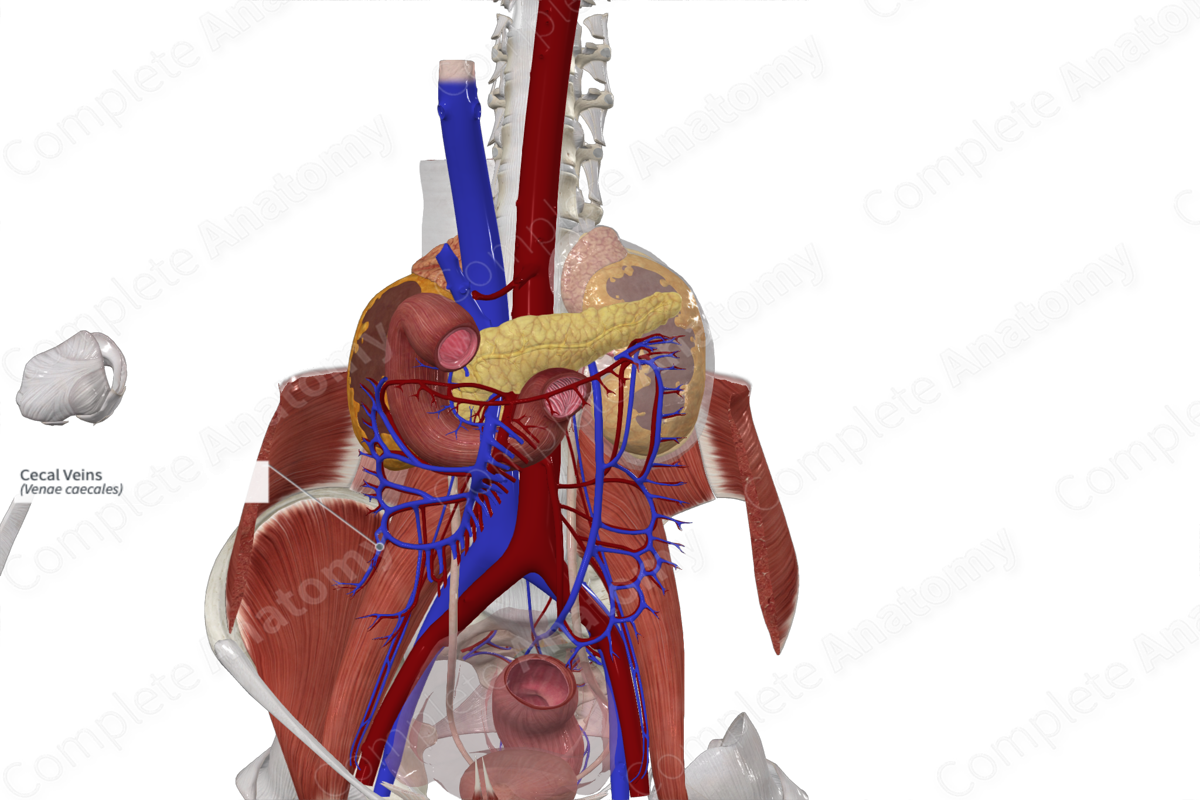
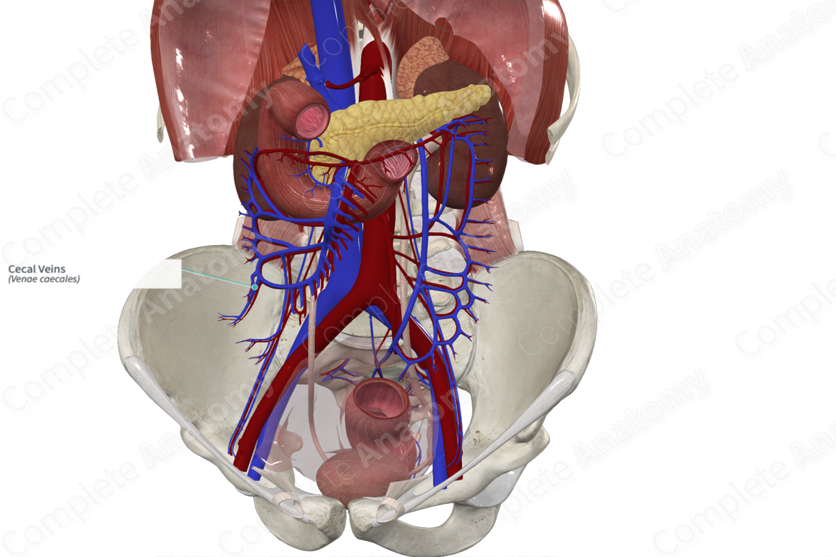
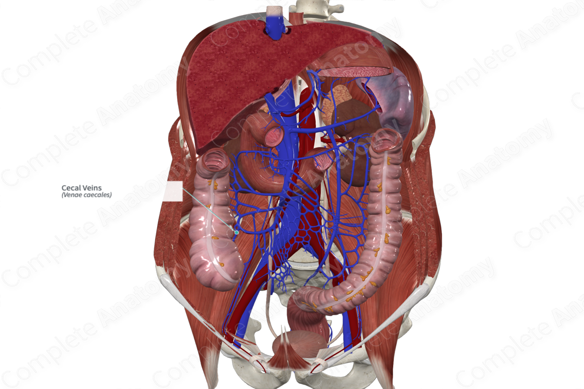
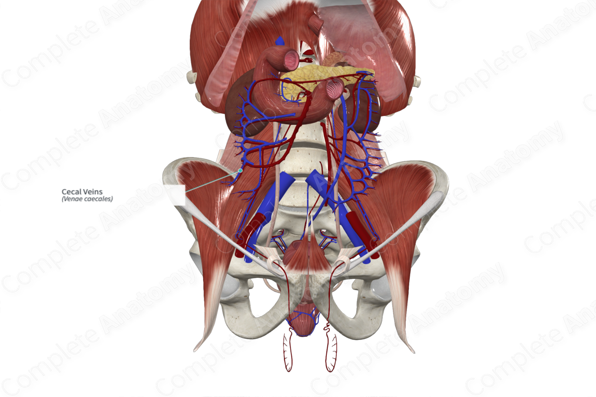
Origin: Along the surface of the cecum.
Course: Travels from inferolateral to superomedial to join the appendicular veins as the ileocolic vein and ultimately the superior mesenteric vein.
Tributaries: None.
Drainage: Cecum.
Origin
The cecal veins (anterior and posterior) originate along the surface of the cecum near the ileocecal junction.
Course
The cecal veins travel superomedially from the cecum (lower right quadrant) to merge with the appendicular veins as the ileocolic vein, which will then drain into the superior mesenteric vein.
Tributaries
There are no named tributaries.
Structures Drained
The cecal veins drain the cecum.
List of Clinical Correlates
- Appendectomy
References
Standring, S. (2016) Gray's Anatomy: The Anatomical Basis of Clinical Practice. Gray's Anatomy Series 41 edn.: Elsevier Limited.
Learn more about this topic from other Elsevier products

