Quick Facts
Origin: Continuation of aortic arch.
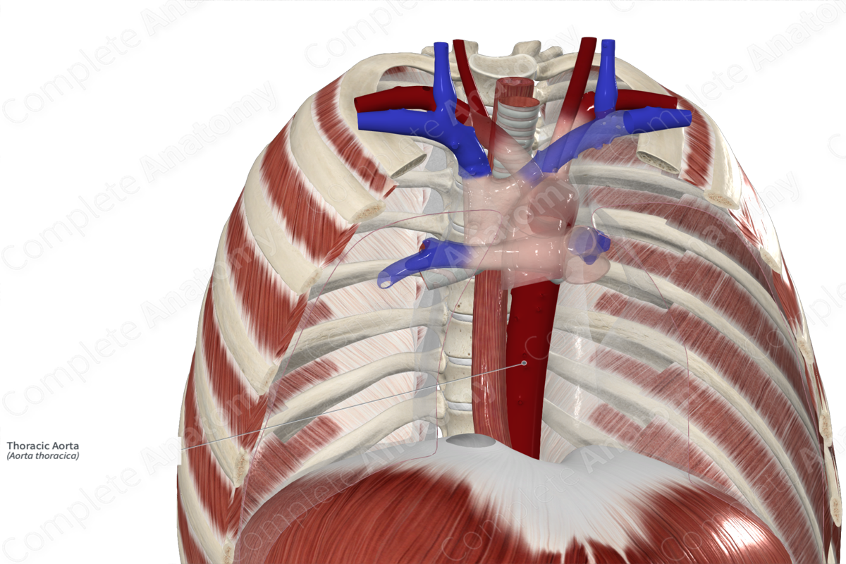
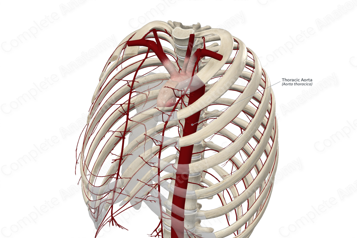
Course: Descends in the posterior mediastinum between the fourth and twelfth thoracic vertebrae.
Branches: Third to eleventh posterior intercostal, subcostal, esophageal, bronchial, superior phrenic, mediastinal, and pericardial arteries.
Supplied Structures: Thoracic viscera, thoracic wall, and respiratory diaphragm.
Origin
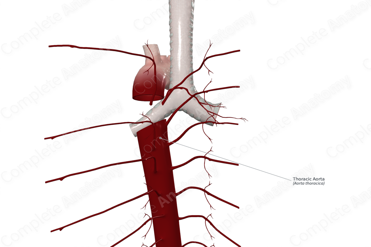
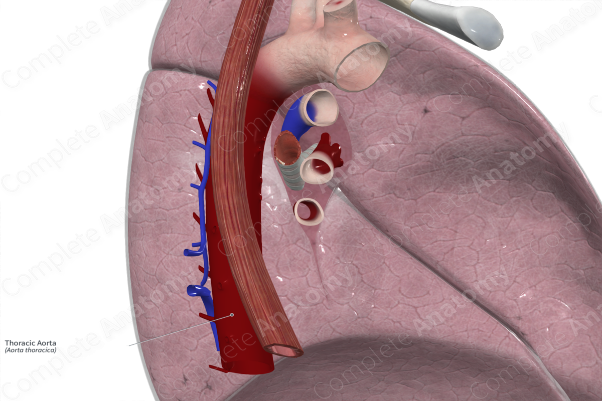
The thoracic aorta is continuous with the aortic arch at the level of the lower border of the fourth thoracic vertebra, to the left of the midline.
Course
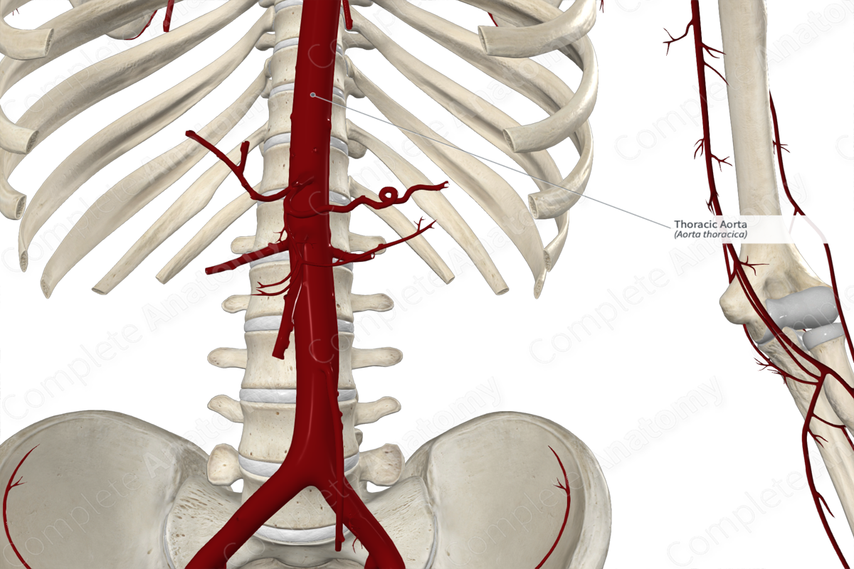
From the lower border the fourth thoracic vertebra, the thoracic aorta descends within the posterior mediastinum. It gradually moves towards the midline and terminates as it passes through the aortic hiatus of the respiratory diaphragm at the level of the twelfth thoracic vertebra.
Branches
The arterial branching of the thoracic aorta tends to arise and follow three vascular planes.
- Anterior plane gives rise to unpaired visceral branches that supply the gut and its derivatives. These include the esophageal arteries which may vary in number.
- Lateral plane gives rise to paired visceral branches that supply the viscera independent of the gut. These arteries are the bronchial arteries.
- Posterolateral plane gives rise to paired parietal branches that supply the body wall. These arteries are the nine posterior intercostal arteries and the subcostal arteries that supply the intercostal (except the first and second) and subcostal spaces.
Some branches of the thoracic aorta do not follow this rule. The superior phrenic arteries are paired parietal branches that pass anterolaterally over the superior surface of the respiratory diaphragm. The pericardial and mediastinal arteries are unpaired visceral branches that arise anteriorly, however, do not pass to the gut.
Supplied Structures
The thoracic aorta and its branches provide an arterial supply to the thoracic viscera, thoracic wall, and respiratory diaphragm.
List of Clinical Correlates
- Aortic dissection
- Atherosclerosis
Learn more about this topic from other Elsevier products

