Description
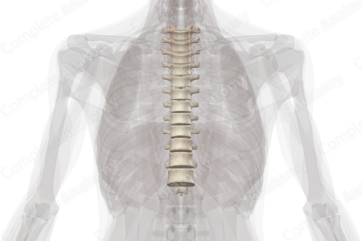
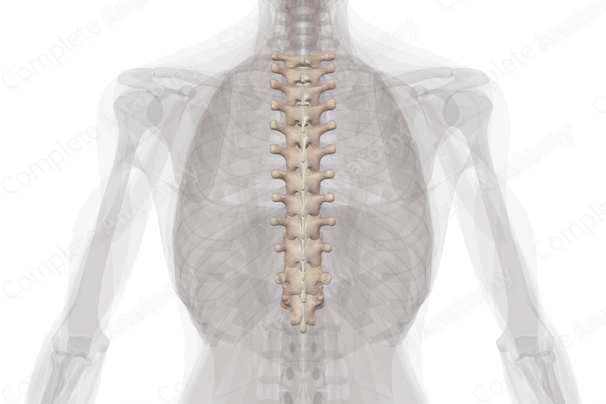
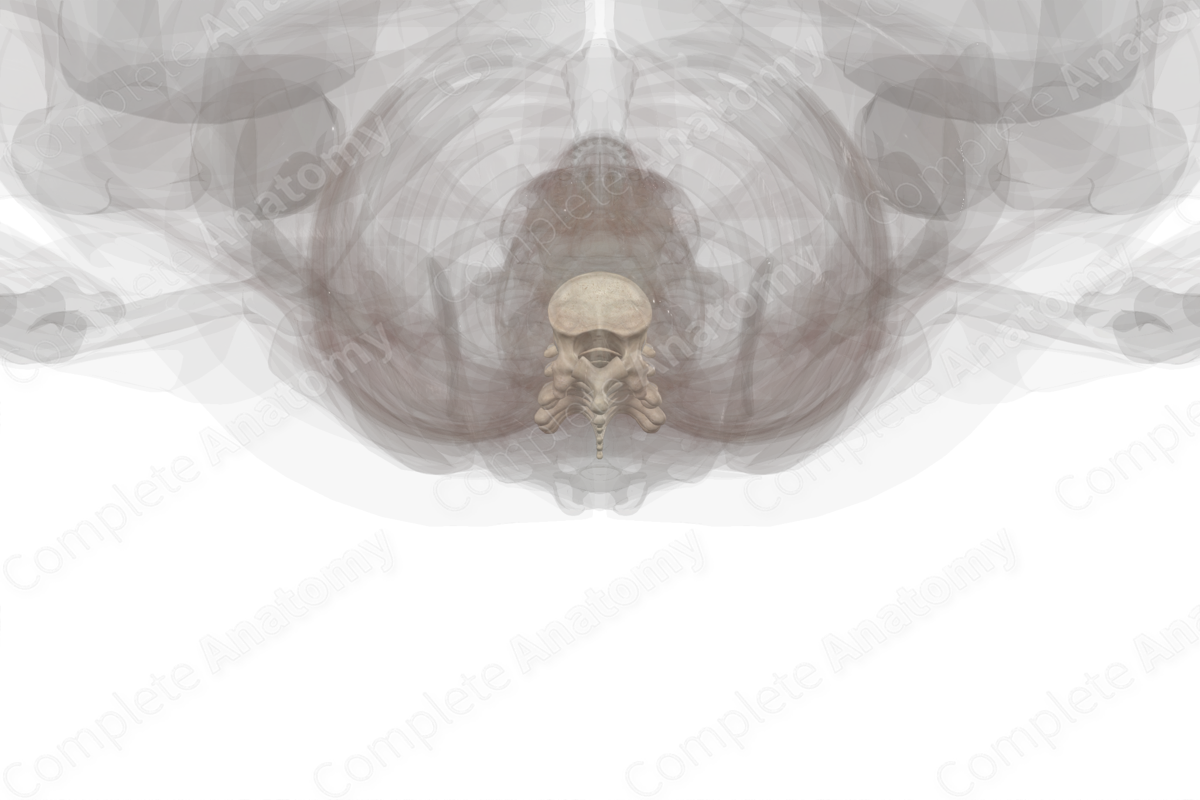
The thoracic vertebrae are one of the five regional groups of the vertebral column. They are found along the posterior aspect of the thorax and are located between the cervical and lumbar vertebrae. There are twelve thoracic vertebrae, which become progressively larger from superior to inferior. This progressive increase in size is necessary to support the progressive increase in body weight that is placed on each successive vertebra. The thoracic vertebrae are unfused and articulate with adjacent vertebrae via:
- intervertebral discs, forming intervertebral symphysis joints;
- articular facets, forming zygapophyseal joints.
The general characteristic features of thoracic vertebrae include:
- vertebral bodies that appear heart-shaped when viewed from above and consist of costal facets;
- vertebral foramina that appear circular when viewed from above;
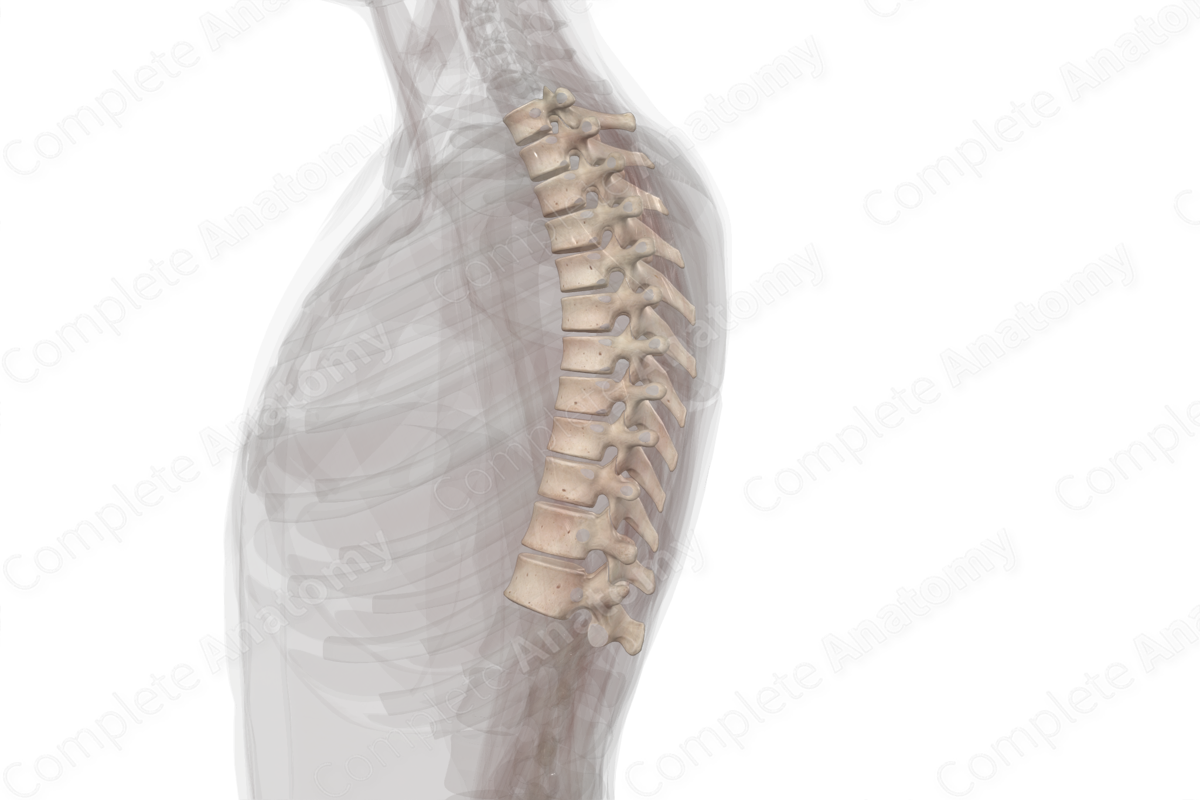
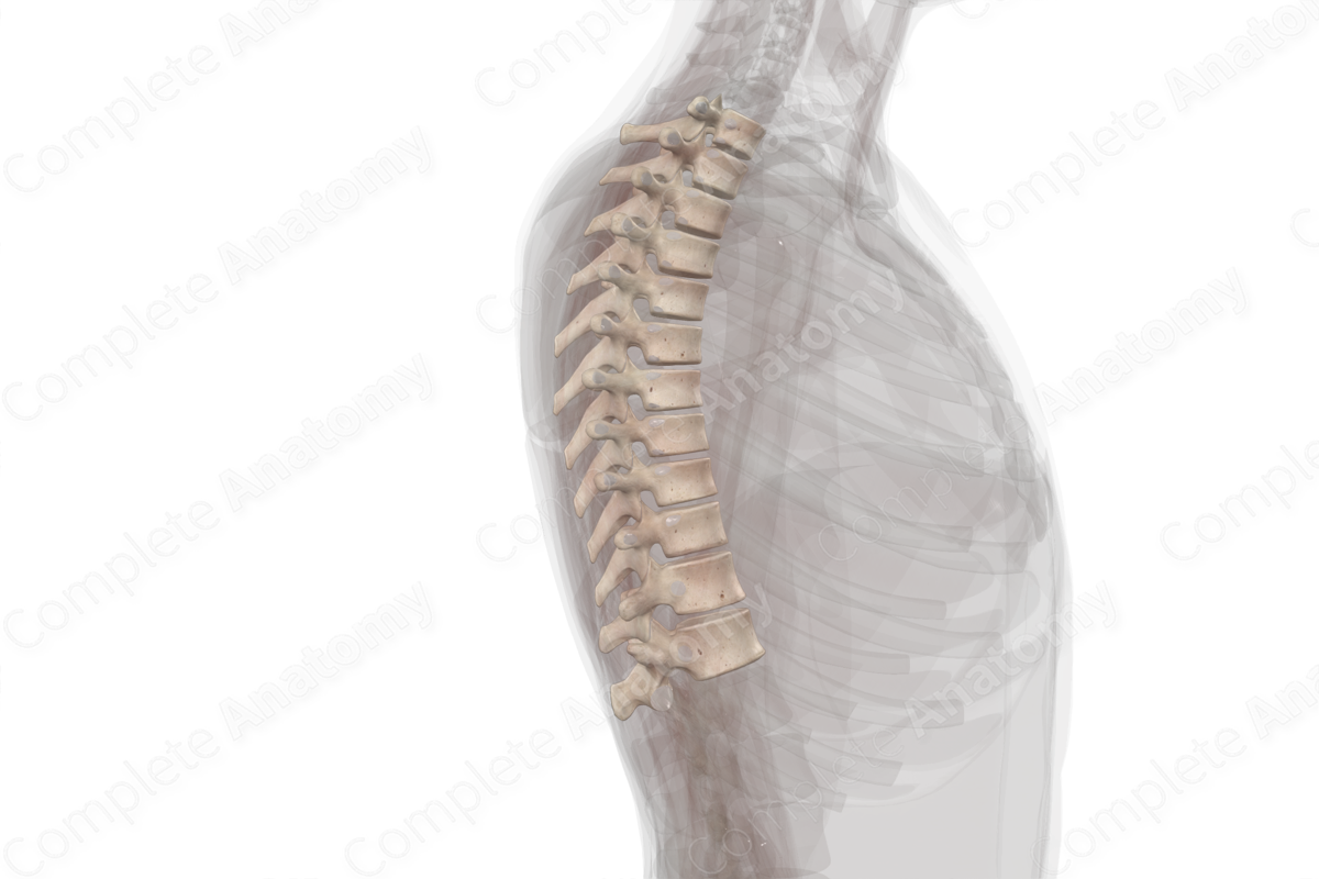
- spinous processes that are short, thick, tapered, and obliquely oriented in the posteroinferior direction;
- transverse processes that are long, thin, and have expanded lateral ends, with most of them consisting of costal facets;
- superior articular processes that consist of articular facets that face posteriorly and slightly laterally;
- inferior articular processes that consist of articular facets that face anteriorly and slightly medially;
- costal facets that articulate with the ribs at costovertebral joints.
Related parts of the anatomy
List of Clinical Correlates
- Excessive thoracic kyphosis/hunch back
- Spinal stenosis
- Intervertebral disc herniation
- Scoliosis
- Spondylosis
- Spondylolisthesis
- Spondylolysis
- Compression fracture
- Butterfly vertebra
- Spina bifida
Learn more about this topic from other Elsevier products

