Quick Facts
Location: Left side of the thoracic cavity.
Arterial Supply: Left bronchial arteries.
Venous Drainage: Left bronchial veins.
Innervation: Pulmonary plexus.
Lymphatic Drainage: Superficial and deep lymphatic plexuses, bronchopulmonary and intrapulmonary lymph nodes.
Related parts of the anatomy
Structure
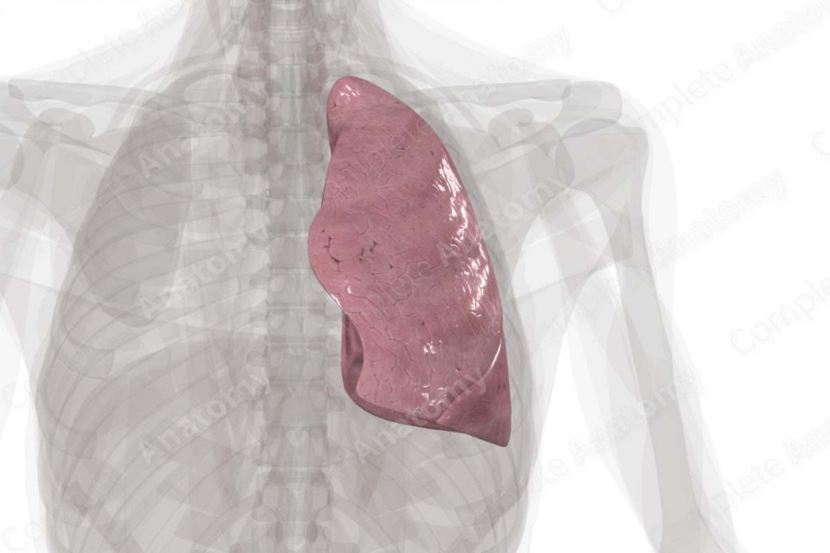
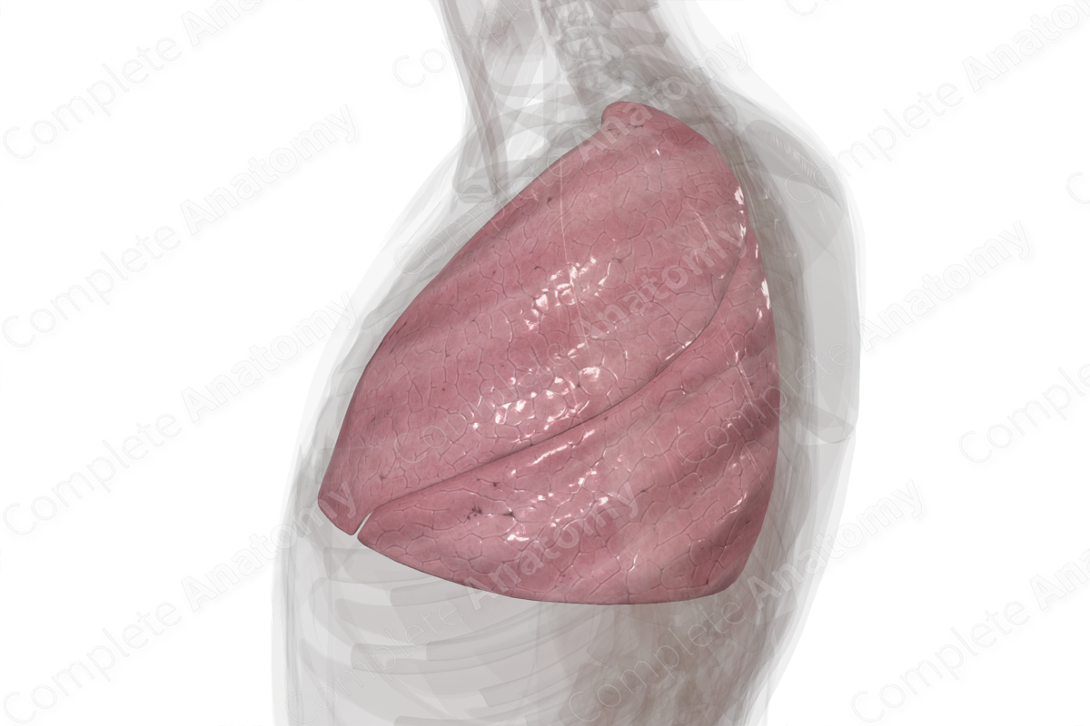
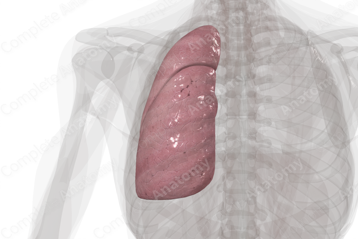
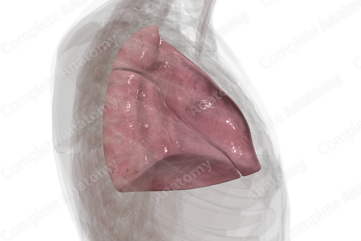
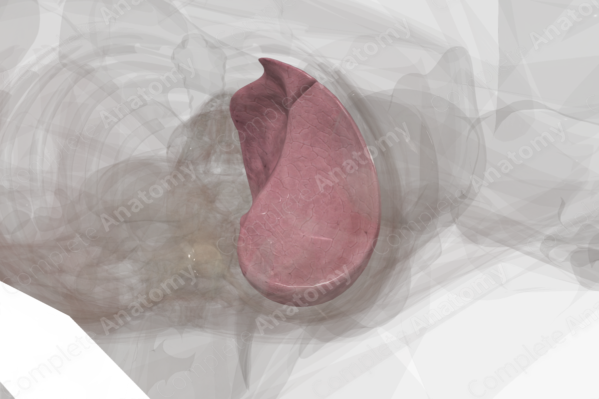
The left lung consists of lung parenchyma and supportive connective tissue. It is subdivided into two lobes; the superior and inferior lobes. These lobes are separated by the interlobar fissure known as the oblique fissure.
The lungs are conical in shape, with a superiorly located apex and an inferiorly located base. Each lung has three surfaces:
- medially located mediastinal surface;
- laterally located costal surface;
- inferiorly located diaphragmatic surface (or the base of the lung).
The three lung surfaces are separated by three borders:
- the anterior border separates the anterior end of the mediastinal surface from the costal surface;
- the posterior border separates the posterior end of the mediastinal surface from the costal surface;
- the inferior border separates the inferior ends of both the mediastinal and costal surfaces from the diaphragmatic surface.
Located along the mediastinal surface of each lung is the hilum, the site where the arteries, veins, nerves, and bronchi enter the lung. The collective term for these structures that enter the lung through the hilum is the “root” of the lung.
On the left, due to the cardiac notch, the lung is pushed laterally at the level of the fourth costal cartilage (approximately 3.5 cm) and curves inferomedially to the sixth costal cartilage. This thin inferomedial projection is called the lingula. It enters the costomediastinal recess during respiration (Moore, Dalley and Agur, 2013).
Anatomical Relations
The apex of each lung is located:
- posterior to the subclavian artery, internal thoracic artery, and scalenus anterior muscle;
- anterior to the neck of the first rib, ventral ramus of first thoracic nerve, first posterior intercostal artery, first posterior intercostal vein, and the sympathetic chain;
- superior to the medial one third of clavicle, by approximately 2.5 cm.
The diaphragmatic surface (or the base) of each lung is located superior to the diaphragm. The right hemidiaphragm separates the right lung from the liver, while the left hemidiaphragm separates the left lung from the spleen and fundus of stomach. The costal surface of each lung lies deep to the ribs and innermost intercostal muscles. The mediastinal surface of each lung lies lateral to the mediastinum. The right lung lies lateral to the right atrium, superior vena cava, right brachiocephalic, azygos vein, trachea, esophagus, and the right phrenic and vagus nerves. The left lung lies lateral to the left ventricle, aorta, left subclavian artery, left brachiocephalic vein, trachea, esophagus, and left phrenic and vagus nerves.
Cadaveric lung impressions demonstrate the differences in the mediastinal relations. On the left lung, there is a deep impression from the left ventricle which forms the cardiac notch. The aortic arch, the left subclavian artery, and the descending thoracic aorta are also identified. The esophagus may leave an impression on the lower portion of the left lung. It is only the cardiac impressions that are seen during surgery as the others are artifacts of cadaveric fixation (Moore, Dalley and Agur, 2013).
Hilum of Left Lung
The pulmonary hilum sits on the medial surface of the lungs and connects the lungs to the heart and the trachea. It sits at the level of the fifth to seventh thoracic vertebral bodies (T5—T7). It is formed by several structures that are entering or exiting the lung, including the:
- principal bronchus;
- pulmonary artery,
- superior and inferior pulmonary veins;
- bronchial vessels;
- pulmonary plexus;
- lymph vessels and bronchopulmonary lymph nodes.
These structures are enveloped in loose connective tissue which in turn is encapsulated by a pleural sleeve.
On both sides, the phrenic nerve, pericardiacophrenic vessels, and anterior pulmonary plexus sit anterior to the hilum. Posterior to the hilum are the vagus nerve and the posterior pulmonary plexus. The pulmonary ligament sits inferiorly.
The right hilum lies posterior to the superior vena cava and the right atrium; however, the left hilum sits inferior to the arch of the aorta and in front of the descending aorta.
The majority of structures are arranged so that the superior pulmonary vein sits anteriorly; the inferior pulmonary vein sits inferiorly; the pulmonary artery and principal bronchus are superior and posterior to the veins, and the bronchial vessels are most posterior.
The position of the pulmonary artery and main bronchus are different in the left and right lungs. In the right, the pulmonary artery sits anterior to the main bronchus, but in the left lung the pulmonary artery sits superior to the main bronchus.
In cadaveric dissection there may appear to be additional structures; for example, the main bronchus may already be divided into lobar bronchi.
Function
The lungs are involved in respiration, which is the process of gas exchange between the atmospheric air and the blood. The lungs consist of lung parenchyma and supportive connective tissue. The lung parenchyma is the functional tissue of the lungs, where gas exchange occurs with pulmonary capillaries; it is composed of alveolar ducts and alveoli.
Arterial Supply
The right and left bronchial arteries are distal branches of the thoracic aorta. They supply oxygenated blood to the tissues of the tracheobronchial tree and the visceral pleura.
Venous Drainage
The bronchial veins that drain the more proximal regions of the tracheobronchial tree drain into the heart via azygos and accessory hemiazygos veins (i.e., via the systemic circulation). The bronchial veins that drain the more distal regions of the tracheobronchial tree form anastomoses with pulmonary veins and drain back to the heart via these veins (i.e., via the pulmonary circulation).
Innervation
The nerve supply to the lungs and visceral pleurae is via the pulmonary plexus. The pulmonary plexus provides autonomic (i.e., visceral) innervation to these structures, and it consists of both parasympathetic and sympathetic fibers.
- Parasympathetic stimulation within the respiratory system promotes bronchoconstriction, vasodilation of pulmonary blood vessels, and secretion from glands within the walls of the tracheobronchial tree.
- Sympathetic stimulation promotes bronchodilation, and vasoconstriction of pulmonary blood vessels.
Lymphatic Drainage
The lymphatic drainage system within the lungs can be divided into two plexuses; the superficial plexus and the deep plexus.
- The superficial lymphatic plexus (or subpleural plexus) is located within the lung parenchyma adjacent to the visceral pleura. This plexus provides lymphatic drainage to the superficial regions of the lungs and the distal ends of the tracheobronchial tree. It drains into the bronchopulmonary lymph nodes.
- The deep lymphatic plexus is located within the walls of the proximal tracheobronchial tree and the lung parenchyma adjacent to it. This plexus drains these areas, and, in turn, drains into the intrapulmonary lymph nodes.
List of Clinical Correlates
- Lung cancer
- Pulmonary collapse (atelectasis)
- Lung resection
References
Moore, K. L., Dalley, A. F. and Agur, A. M. R. (2013) Clinically Oriented Anatomy. Clinically Oriented Anatomy 7th edn.: Wolters Kluwer Health/Lippincott Williams & Wilkins.
