Quick Facts
Location: Left side of the thoracic cavity.
Arterial Supply: Left bronchial arteries.
Venous Drainage: Left bronchial veins.
Innervation: Pulmonary plexus.
Lymphatic Drainage: Superficial and deep lymphatic plexuses, intrapulmonary and bronchopulmonary lymph nodes.


Related parts of the anatomy
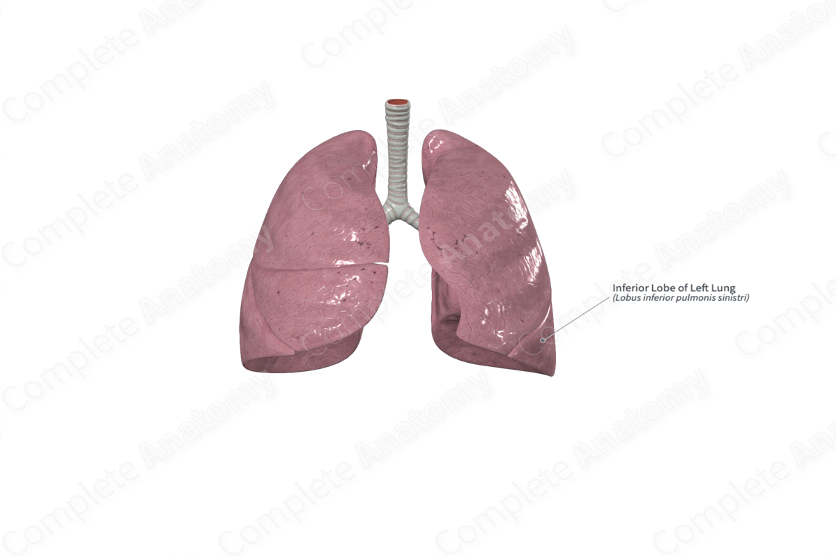
Structure
The left lung has two lobes, the superior and inferior lobes, which are separated by the oblique fissure. The inferior lobe forms the base of the left lung and a portion of the costal and mediastinal surfaces. The superior lobe is anterosuperior to the inferior lobe and makes up the apex and the majority of the mediastinal and costal surfaces.
Like the inferior lobe of the right lung, the inferior lobe of the left lung is subdivided into five bronchopulmonary segments; the superior, medial basal, lateral basal, anterior basal, and posterior basal bronchopulmonary segments. Each of these bronchopulmonary segments has its own segmental bronchus and segmental artery.
Anatomical Relations
The inferior lobe of the left lung sits inferior to the oblique fissure. This fissure begins on the mediastinal surface, at the posterosuperior margin of the hilum. It courses in a superoposterior direction to the posterior border of the lung, 6 cm inferior to the apex. It descends anteriorly along the costal surface as far as the anterior portion of the inferior border. It then continues onto the mediastinal surface and extends as far as the inferior border of the hilum.
The inferior lobe of the left lung sits over the left hemidiaphragm. Therefore, the inferior lobe on the left is in close proximity to the fundus of the stomach and the spleen.
Function
The lungs are involved in respiration, which is the process of gas exchange between the atmospheric air and the blood. The lungs consist of lung parenchyma and supportive connective tissue. The lung parenchyma is the functional tissue of the lungs, where gas exchange occurs with pulmonary capillaries; it is composed of alveolar ducts and alveoli.
Arterial Supply
The right and left bronchial arteries are distal branches of the thoracic aorta. They supply oxygenated blood to the tissues of the tracheobronchial tree and the visceral pleura.
Venous Drainage
The bronchial veins that drain the more proximal regions of the tracheobronchial tree drain into the heart via azygos and accessory hemiazygos veins (i.e., via the systemic circulation). The bronchial veins that drain the more distal regions of the tracheobronchial tree form anastomoses with pulmonary veins and drain back to the heart via these veins (i.e., via the pulmonary circulation).
Innervation
The nerve supply to the lungs and visceral pleurae is via the pulmonary plexus. The pulmonary plexus provides autonomic (i.e., visceral) innervation to these structures, and it consists of both parasympathetic and sympathetic fibers.
- Parasympathetic stimulation within the respiratory system promotes bronchoconstriction, vasodilation of pulmonary blood vessels, and secretion from glands within the walls of the tracheobronchial tree.
- Sympathetic stimulation promotes bronchodilation, and vasoconstriction of pulmonary blood vessels.
Lymphatic Drainage
The lymphatic drainage system within the lungs can be divided into two plexuses; the superficial plexus and the deep plexus.
- The superficial lymphatic plexus (or subpleural plexus) is located within the lung parenchyma adjacent to the visceral pleura. This plexus provides lymphatic drainage to the superficial regions of the lungs and the distal ends of the tracheobronchial tree. It drains into the bronchopulmonary lymph nodes.
- The deep lymphatic plexus is located within the walls of the proximal tracheobronchial tree and the lung parenchyma adjacent to it. This plexus drains these areas, and, in turn, drains into the intrapulmonary lymph nodes.
List of Clinical Correlates
- Lung cancer
- Pulmonary collapse (atelectasis)
- Lung resection
