Quick Facts
The dura mater is the outermost, toughest, and most fibrous of the three membranes (meninges) covering the brain and spinal cord (Dorland, 2011).
Structure and/or Key Features
There are three layers that make up the meninges, the dura, arachnoid, and pia mater. The meningeal layers can also be divided according to the stage of their development, including the leptomeninges (arachnoid and pia mater) and pachymeninx (dura mater).
The dura mater is a dense fibrous layer, composed mostly of tightly packed fascicles of collagen in a laminae configuration. It lies external to the arachnoid and is separated from the arachnoid by the subdural space.
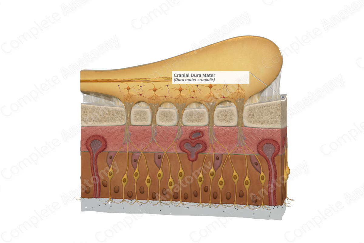
The cranial dura mater has two layers, the external endosteal layer and the internal meningeal layer. The endosteal layer of the cranial dura mater is adherent with the inner surface of the calvaria, particularly at the sutures, the cranial base, and in the vicinity of the foramen magnum. The endosteal layer is continuous with the periosteum on the outer aspect of the calvaria at the cranial foramina, while the meningeal layer is continuous with the spinal dura at the foramen magnum. The two layers are inseparable, except at points where the dural sinuses and dural reflections arise. The meningeal layer of the dura mater is closely associated with the arachnoid and both layers are fused at points where veins course from the brain into the venous sinuses (Moore, Dalley and Agur, 2013; Standring, 2016).
Dural reflections, or infoldings, are formed by the inner meningeal layer of the dura at points where it separates from the external endosteal layer. The dural reflections include the falx cerebri, falx cerebelli, tentorium cerebelli, and diaphragma sellae. These reflections divide the cranial cavity into various compartments (Moore, Dalley and Agur, 2013).
The falx cerebri is shaped like a crescent. It narrows anteriorly towards the point where it attaches to the crista galli, and widens posteriorly to merge with the tentorium cerebelli. It is the largest of the four dural reflections and is inhabited in the longitudinal fissure between the left and right cerebral hemispheres. The inferior edge of the falx is free and houses the inferior sagittal sinus. The falx cerebri terminates by continuing as the tentorium cerebelli (Moore, Dalley and Agur, 2013).
The falx cerebelli is a small vertical dural reflection that is situated in the posterior cranial fossa, inferior to the tentorium cerebelli. It slightly separates the two cerebral hemispheres. Posteriorly, it is attached to the internal occipital crest and inhabits the occipital sinus (Standring, 2016)
The diaphragma sellae, the smallest of the four dural reflections, is a spherical horizontal sheet that hangs between the clinoid processes and forms a roof over the sella turcica, encircling the pituitary gland.
The tentorium cerebelli is the second-largest dural reflection and shaped like a crescent. It is situated between the cerebellum and the occipital lobes. It separates the cranial cavity into supra- and infra-tentorial segments where the forebrain and hindbrain are situated, respectively. The falx cerebri affixes itself to the tentorium cerebelli and props it up, hence the similarity to a tent. It connects to the sphenoid bone (rostrally), the petrous portion of the temporal bone (rostrolaterally), the occipital bone, and the parietal bone (posterolaterally) (Moore, Dalley and Agur, 2013).
Anatomical Relations
The dura mater forms the external sheet of the meninges and is separated from the arachnoid by the subdural space.
Function
The principal function of meningeal layers is to protect and provide support to the brain and spinal cord. The dura mater is thickest and most resilient of the three meningeal layers; hence it shields the brain and spinal cord from mechanical trauma.
The dura mater encircles and supports the dural venous sinuses that convey blood from the brain to the heart. It is also responsible for sealing the central nervous system to ensure cerebrospinal fluid is kept within.
Clinical Correlates
—Subdural hemorrhage
—Epidural hemorrhage
—Epidural injections
References
Dorland, W. (2011) Dorland's Illustrated Medical Dictionary. 32nd edn. Philadelphia, USA: Elsevier Saunders.
Moore, K. L., Dalley, A. F. and Agur, A. M. R. (2013) Clinically Oriented Anatomy. Clinically Oriented Anatomy 7th edn.: Wolters Kluwer Health/Lippincott Williams & Wilkins.
Standring, S. (2016) Gray's Anatomy: The Anatomical Basis of Clinical Practice. Gray's Anatomy Series 41st edition: Elsevier Limited.
