Quick Facts
Supporting cells are cells that serve to provide support and protection and perhaps contribute to the nutrition of principal or other cells of certain organs; included are cells in the membranous labyrinth of the internal ear, olfactory epithelium, taste buds, and seminiferous tubules (Sertoli cells) (Dorland, 2011).
Structure and/or Key Features
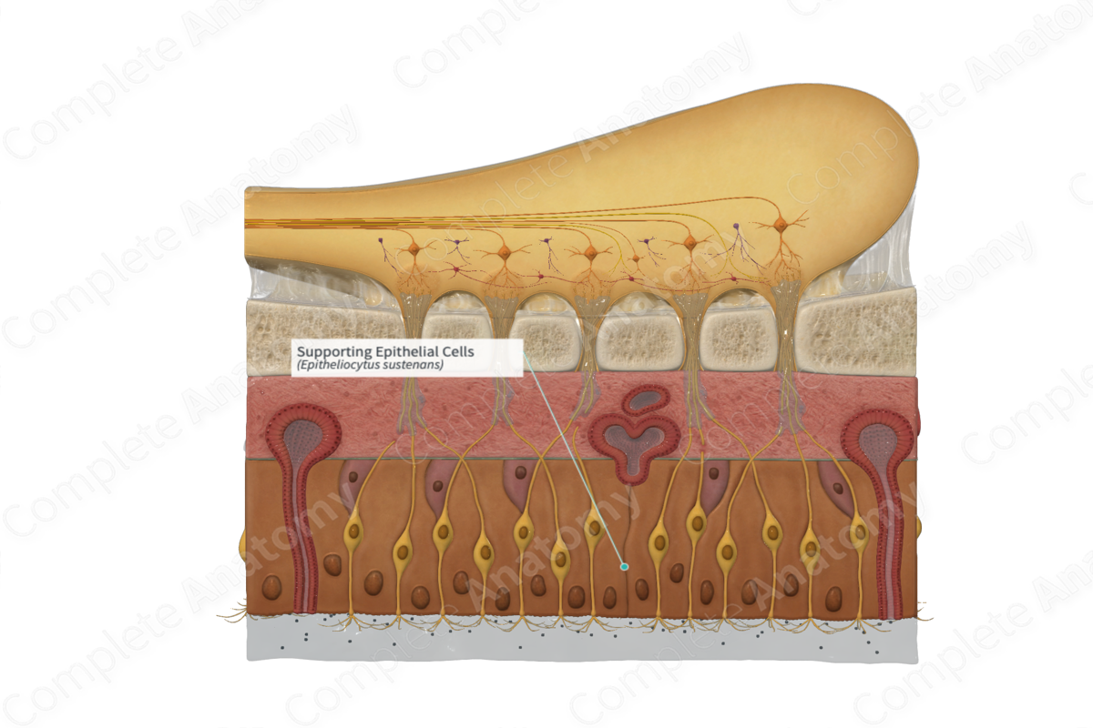
The olfactory epithelium is made up of various cell types such as basal cells, olfactory sensory neurons, and the supporting epithelial cells (Rao, Vemuri and Carpenter, 2007).
The supporting cells in the olfactory epithelium can be likened to the glial cells in the nervous system. They are non-neural cells that are located in the apical layer of the olfactory epithelium (Cheok and Karunanayaka, 2018). There are two types of supporting cells, the sustentacular and microvillar cells.
The microvillar cells take up a superficial location within the olfactory epithelium. They are electron-lucent and shaped like flasks. At the apex of microvillar cells, there are microvilli that extend into the mucus layer that overlies the olfactory epithelium. The thickness of microvillar cells are about one tenth that of olfactory sensory neurons (Standring, 2016).
The sustentacular cells are columnar cells that divide and partly envelope the olfactory sensory neurons. There are many microvilli situated on the apical surface of the sustentacular cells. These apical microvilli extend into the mucus layer overlying the olfactory epithelium where they are interconnected with the cilia of the olfactory sensory neurons. Desmosomes connect adjacent sustentacular cells near the epithelial surface, while tight junctions connect sustentacular cells and olfactory sensory neurons at the epithelial surface. These bonds aid in the stability of the olfactory epithelium (Standring, 2016).
Function
There are two types of supporting cells, the sustentacular cells and microvillar cells.
The sustentacular cells provide metabolic and physical support for the olfactory epithelium. The physical support is as a result of the bonds formed by desmosomes and tight junctions between adjacent sustentacular cells, as well as between sustentacular cells and olfactory sensory neurons (Cheok and Karunanayaka, 2018; Standring, 2016).
The function of microvillar cells is not well known. However, their apical microvilli increase the surface area for absorption.
References
Cheok, A. D. and Karunanayaka, K. (2018) Virtual Taste and Smell Technologies for Multisensory Internet and Virtual Reality. Human–Computer Interaction Series: Springer International Publishing.
Dorland, W. (2011) Dorland's Illustrated Medical Dictionary. 32nd edn. Philadelphia, USA: Elsevier Saunders.
Rao, M. S., Vemuri, M. C. and Carpenter, M. (2007) Neural Development and Stem Cells. Contemporary Neuroscience: Humana Press.
Standring, S. (2016) Gray's Anatomy: The Anatomical Basis of Clinical Practice. Gray's Anatomy Series 41st edition: Elsevier Limited.
