Quick Facts
Origin: Ophthalmic nerve.
Course: Runs from the superior orbital fissure into the common tendinous ring, and anteriorly along the medial aspect of the orbit.
Branches: Anterior and posterior ethmoidal nerves, infratrochlear nerve, long ciliary nerve, and branch to ciliary ganglion.
Supply: Sensory: conveys sensation from the skin of the dorsum of the nose and medial eyelids, the medial conjunctiva, the membranes of the ethmoidal air cells and sphenoid sinus, and portions of the nasal cavity; Sympathetic: fibers join the nasociliary nerve to reach the eye and the dilator pupillae muscle.


Related parts of the anatomy
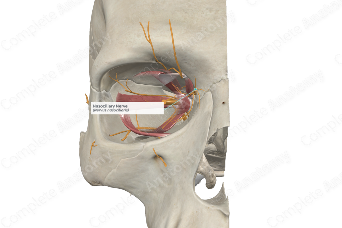
Origin
The nasociliary nerve is a branch of the ophthalmic nerve that originates just proximal to or within the superior orbital fissure.
Course
The nasociliary nerve runs through the superior orbital fissure, traveling within the common tendinous ring and passing between the proximal head of the lateral rectus muscle. It runs obliquely forward through the orbit, crossing over the optic nerve, deep to the superior rectus muscle, and then continues forward between the superior oblique and the inferior rectus muscles on the medial side of the orbit. Along the way, it gives off branches and terminates as either the anterior ethmoidal nerve or the infratrochlear nerve.
Branches
The nasociliary nerve gives rise to a number of branches.
—The anterior and posterior ethmoidal nerves penetrate the ethmoid bone, with the anterior nerve re-emerging below the nasal bone.
—The long ciliary nerves run to the posterior sclera of the eye.
—The branch to the ciliary ganglion runs to the ciliary ganglion and then on to the posterior sclera of the eye via the short ciliary nerves.
—The infratrochlear nerve runs anteriorly on the superior edge of the medial rectus muscle, inferior to the trochlea, and exits the orbit medially, just inferior to the supratrochlear nerve.
Supplied Structures
The nasociliary nerve is largely a sensory nerve that conveys general sensation from the nasal and optic regions of the face, but also has a role in transmitting sympathetic innervation to the dilator pupillae muscle of the eye.
The sensory branches of the nasociliary nerve and their targets are as follows.
—The posterior ethmoidal nerve innervates the mucosa of the sphenoid sinus and posterior ethmoidal air cells.
—The anterior ethmoidal nerve innervates the mucosa of the remaining ethmoidal air cells, the anterior surfaces of the nasal septum and lateral nasal wall, and the skin of the dorsum, ala, and apex of the nose.
—The infratrochlear nerve conveys sensation from the skin of the medial eyelids and bridge of the nose, as well as the medial conjunctiva.
—The long ciliary nerve conveys sensation from the more medial surfaces of the eye.
—The branch to the ciliary ganglion conveys sensation from the more lateral surfaces of the eye.
Sympathetic fibers traveling to the dilator pupillae muscle of the eye run with the nasociliary nerve and travel to the eye either via the long ciliary nerves or the communication to the ciliary ganglion and short ciliary nerves.
List of Clinical Correlates
—Pupillary miosis
Learn more about this topic from other Elsevier products

