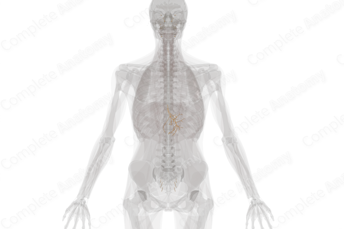
Craniosacral Part of Autonomic Division
Pars craniosacralis divisionis autonomicae
Read moreDescription
The craniosacral part of the autonomic division is the parasympathetic part of the autonomic division, so named because parasympathetic neurons originate in the brainstem (cranial) or from the sacral spinal cord.
Functionally, the parasympathetic part of the autonomic division is associated with states of reduced alertness and increased digestive activity, e.g., constricted pupils, reduced heart rate, reduced blood flow to skeletal muscle, increased blood flow to the digestive tract, and increase in glandular functions associated with digestion. For this reason, the parasympathetic nervous system is often associated with “rest and digest.”
The different parts of the craniosacral part of the autonomic division include:
—preganglionic parasympathetic neurons in the brainstem that contribute to cranial nerves III, VII, IX, and X;
—the ciliary, pterygopalatine, submandibular, and otic ganglia, which are postganglionic parasympathetic ganglia found in the head;
—intramural ganglia, which are small clusters of postganglionic parasympathetic neurons typically found embedded within the walls of the target tissue;
—lateral horn neurons at the S2-S4 spinal cord levels, which are the preganglionic parasympathetic neurons that target the hindgut and pelvis;
—the pelvic splanchnic nerves, which are the only parasympathetic nerves to use the term “splanchnic.”
Related parts of the anatomy
Learn more about this topic from other Elsevier products


