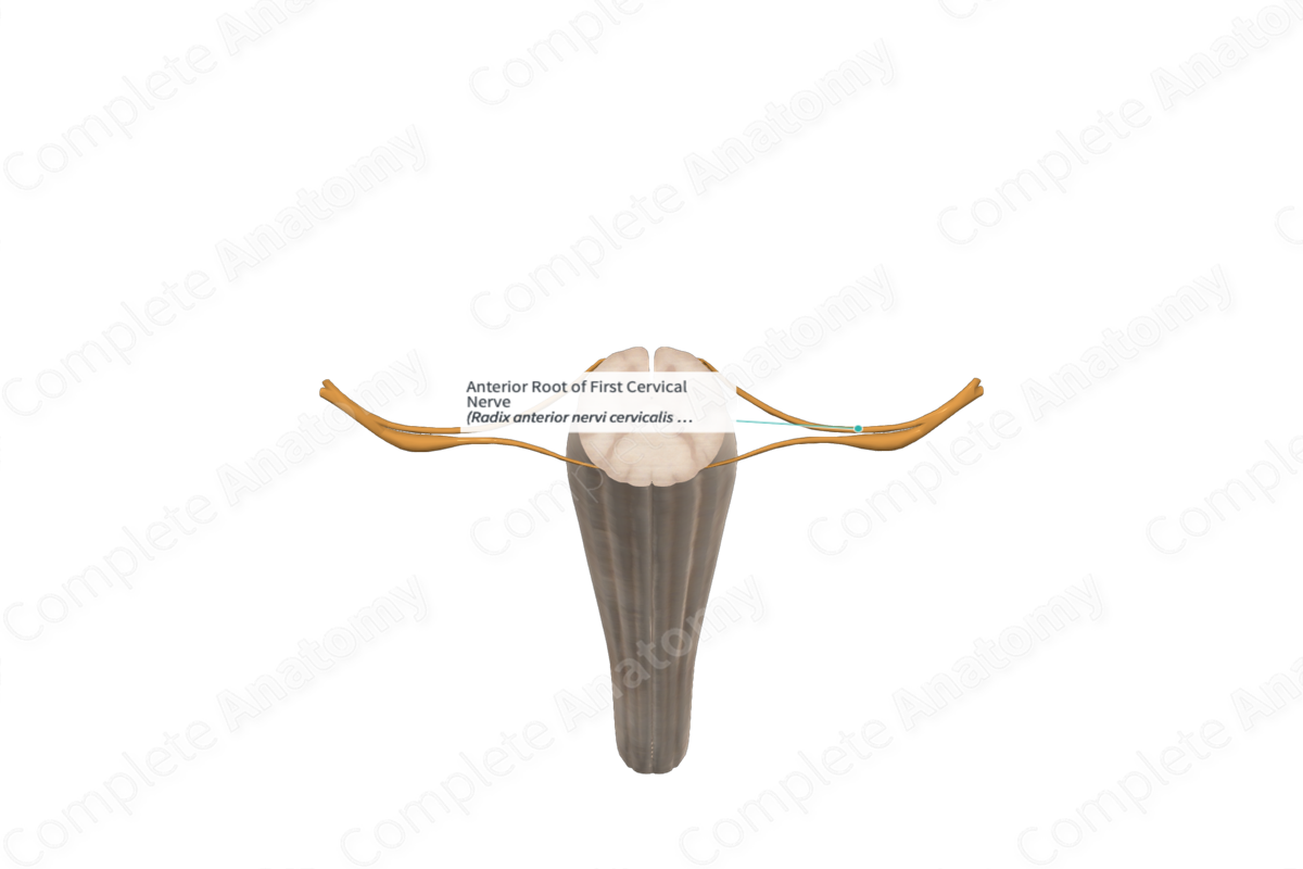
Quick Facts
Origin: Anterolateral sulcus of spinal cord.
Course: Laterally towards the posterior arch of atlas and the occipital bone.
Branches: None.
Supply: Somatic efferent fibers to the first cervical nerve, geniohyoid, infrahyoid, and prevertebral muscles and the muscles bordering the suboccipital triangle.
Related parts of the anatomy
Origin
The anterior root of the first cervical nerve forms from a series of rootlets that emerge from the anterolateral sulcus of the first cervical spinal segment.
Course
The anterior root of the first cervical nerve runs laterally and inferiorly away from the first cervical spinal segment towards a foramen located between the posterior arch of atlas and the occipital bone. Roughly within this foramen, the anterior root merges with the posterior root to form the first cervical nerve.
Size and direction of the spinal roots vary. For instance, the upper cervical roots are short and run horizontally to exit the vertebral canal through the foramen.
Branches
The anterior root of the first cervical nerve merges with the posterior root to form the first cervical nerve and does so without branching.
Supplied Structures
The somatic motor efferents pass through the spinal nerve itself and into either the posterior ramus or the anterior ramus of the first cervical nerve.
Those passing through the anterior ramus contribute to the cervical plexus. It innervates the infrahyoid muscles (sternohyoid, sternothyroid, and superior belly of omohyoid). The thyrohyoid and geniohyoid muscles are also innervated by the anterior ramus of the first cervical nerve via a communicating branch to the hypoglossal nerve.
Fibers that pass through the posterior ramus (or suboccipital nerve) enter the suboccipital triangle to innervate muscles including the rectus capitis posterior major and minor, obliquus capitis superior and inferior, and semispinalis capitis.
Learn more about this topic from other Elsevier products


