Quick Facts
Origin: Third lumbar nerve inside the psoas major muscle.
Course: Contributes to the lumbar plexus.
Branches: Lateral femoral cutaneous, femoral, and obturator nerves.
Supply: Motor innervation to the posterior abdominal wall musculature (psoas major and quadratus lumborum muscles), muscles of the anterior compartment of the thigh (iliacus, pectineus, sartorius, rectus femoris, vastus medialis, vastus intermedius, and vastus lateralis muscles) and various other muscles (adductor brevis, adductor longus, adductor magnus, gracilis, pectineus, and obturator externus muscles). Sensory innervation to the skin of the mons pubis, labium majus, anterior, medial, and lateral thigh regions, down to the level of the knee joint, and skin on the medial side of the leg, ankle, and foot. Moreover, articular branches innervate the hip and knee joints.


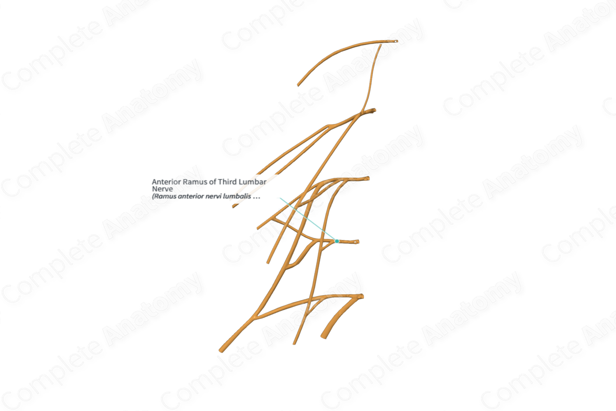
Origin
The anterior (ventral) ramus of the third lumbar nerve originates as one of two branches of the third lumbar nerve. The other branch is the posterior (dorsal) ramus.
Course
The bifurcation of the third lumbar nerve into anterior and posterior rami takes place immediately after its exit from the intervertebral foramen.
The anterior ramus of third lumbar nerve contributes to the formation of the lumbar plexus, which also receives the nerve fibers from the anterior rami of the first, second, and fourth lumbar and the subcostal nerves.
Branches
The anterior ramus of third lumbar nerve is a mixed nerve which contains both somatic efferent (motor) and afferent (sensory) neurons.
The somatic efferent neurons emerge from the anterior gray horn of the L3 segment of the spinal cord. These are lower motor neurons which exit the spinal cord through the anterolateral sulcus, as they travel inside the anterior motor rootlets, and root of L3 spinal segment. They subsequently travel through the third lumbar nerve to enter the anterior ramus before reaching the lumbar plexus. These efferent neurons travel through the muscular branches of the femoral and obturator nerves, to supply motor innervation to various muscles.
Somatic afferent neurons travel through the lateral femoral cutaneous nerve of the thigh, cutaneous nerve branches of the obturator and femoral nerve (medial, intermediate, and saphenous cutaneous nerves), to enter the lumbar plexus. From here onwards, these neurons travel through the anterior ramus of the third lumbar nerve to enter the sensory root and rootlets. The cell bodies of these sensory neurons are located inside the spinal ganglion of the third lumbar nerve. The axons then travel through the posterolateral sulcus to enter the posterior sensory horn of the L3 spinal cord segment.
The anterior ramus of third lumbar nerve is also connected to the sympathetic trunk through the gray communicating branch (gray ramus communicans), which serves as a conduit for the postganglionic sympathetic neurons.
Supplied Structures & Function
The anterior ramus of third lumbar nerve supplies motor innervation to the posterior abdominal wall musculature (psoas major and quadratus lumborum) through the somatic efferent neurons coming directly from the ramus. In addition, these efferent neurons travel through the muscular branches of femoral nerve to innervates muscles of the anterior compartment of the thigh (iliacus, pectineus, sartorius, rectus femoris, vastus medialis, vastus intermedius, and vastus lateralis). Moreover, the efferent neurons from the L3 anterior ramus travel through muscular branches of obturator nerve to innervate muscles, including, adductor brevis, adductor longus, adductor magnus, gracilis, pectineus, and obturator externus.
The somatic afferent neurons from the lateral femoral cutaneous nerve of thighs conduct sensory information from the anterior and lateral thigh region to the level of the knee joint.
The general somatic afferent neurons from the medial, intermediate, and saphenous cutaneous branches of the femoral nerve transmit general sensations from the anterior and medial parts of thigh, skin on the medial side of the leg, ankle, and foot. Some neurons exit from the obturator and femoral nerves to innervate the hip, and knee joints.
Learn more about this topic from other Elsevier products

