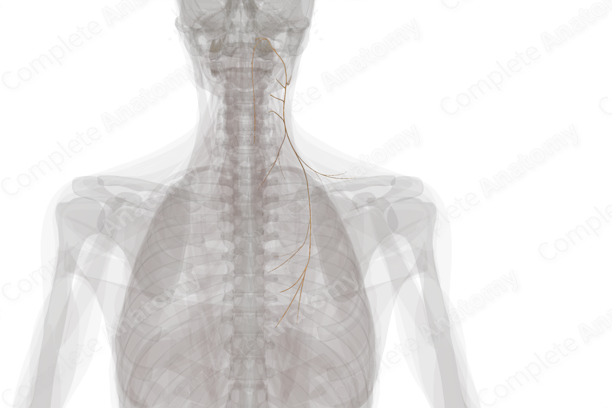
Quick Facts
Origin: Accessory nucleus of cervical spinal cord segments C1-C6.
Course: Ascends into the cranial vault through the foramen magnum, then exits the skull via the jugular foramen. It runs inferior and laterally to the deep surface of the sternocleidomastoid first, and then on to the deep surface of the trapezius.
Branches: Muscular branches.
Supply: Motor innervation of the sternocleidomastoid and trapezius muscles.
Related parts of the anatomy
Origin
The accessory nerve fibers originate in the accessory nucleus of the upper six cervical spinal cord segments (spinal root of accessory nerve). These fibers emerge from the spinal cord laterally.
The accessory nerve also has a cranial component (cranial root of accessory nerve), which is distinct from the fibers of the accessory nerve discussed above. These cranial fibers originate in the medulla oblongata in the same nuclei that give rise to the vagus nerve (Ryan et al, 2007).
Course
The spinal root of the accessory nerve runs superiorly, before ascending into the skull via the foramen magnum. The nerve runs laterally to and then through the jugular foramen to exit the skull, along with the glossopharyngeal and vagus nerves.
The cranial root fibers of the accessory nerve briefly join with and follow the spinal root fibers as they exit the jugular foramen, thus, forming the trunk of the accessory nerve. The fibers originating from the cranial component then quickly dissociate from the spinal fibers and run instead with the vagus nerve, innervating pharyngeal and laryngeal muscles. Functionally and anatomically, the cranial accessory nerve is best considered a portion of the vagus nerve (Ryan et al, 2007). Thus, the fibers originating from the spinal cord are now termed the spinal accessory nerve.
After leaving the skull, the spinal accessory nerve runs inferiorly and posteriorly. It typically crosses in front of the jugular vein on its way to the deep surface of the sternocleidomastoid (SCM) muscle where some fibers terminate. The remainder of the spinal accessory nerve continues obliquely across the posterior triangle, running along the investing facial layer that envelopes the SCM and trapezius muscles. It terminates on the deep surface of the trapezius muscle.
Branches
The spinal accessory nerve gives rise muscular branches to the SCM and trapezius muscle.
Supplied Structures
The spinal accessory nerve is a motor nerve that conveys general somatic efferents to the SCM and trapezius muscles (Kierner et al, 2001).
References
Kierner, A. C., Zelenka, I. & Burian, M. (2001) How do the cervical plexus and the spinal accessory nerve contribute to the innervation of the trapezius muscle? As seen from within using Sihler's stain. Arch Otolaryngol Head Neck Surg, 127(10), 1230-2.
Ryan, S., Blyth, P., Duggan, N., Wild, M. & Al-Ali, S. (2007) Is the cranial accessory nerve really a portion of the accessory nerve? Anatomy of the cranial nerves in the jugular foramen. Anat Sci Int, 82(1), 1-7.
Learn more about this topic from other Elsevier products
Anatomy of the spinal accessory (CN XI) and hypoglossal (CN XII) nerves: Video, Causes, & Meaning

Anatomy of the spinal accessory (CN XI) and hypoglossal (CN XII) nerves: Symptoms, Causes, Videos & Quizzes | Learn Fast for Better Retention!


