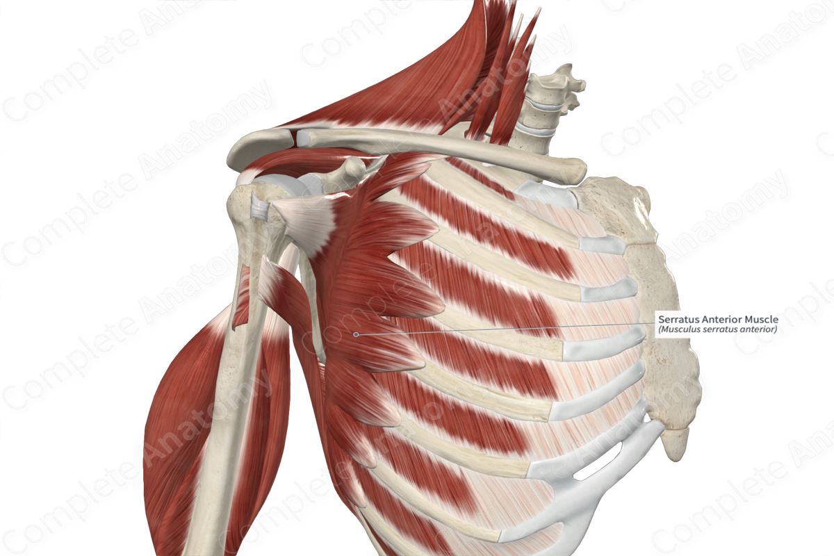
Quick Facts
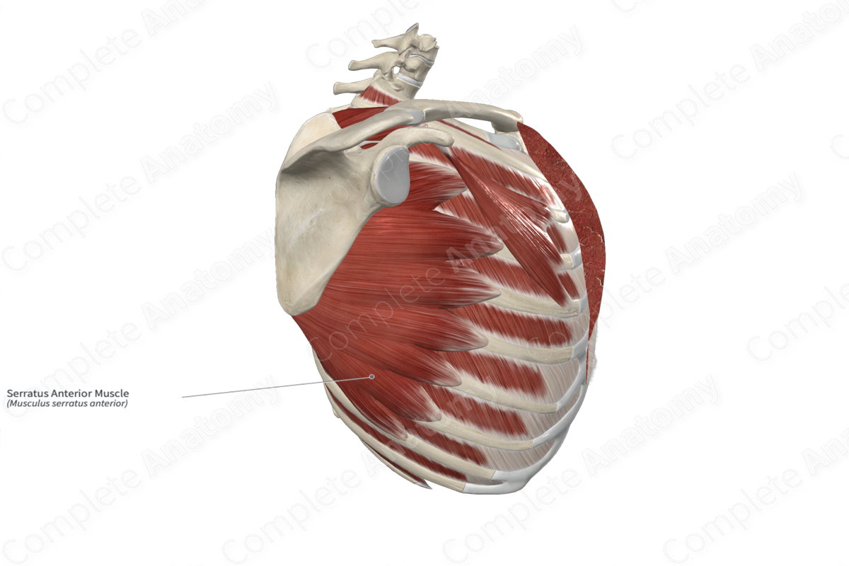
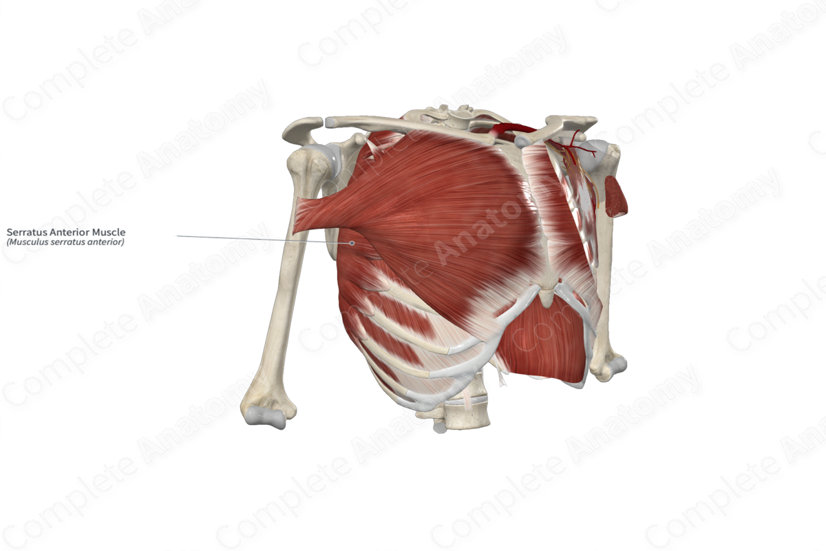
Origin: External surfaces of first to ninth ribs.
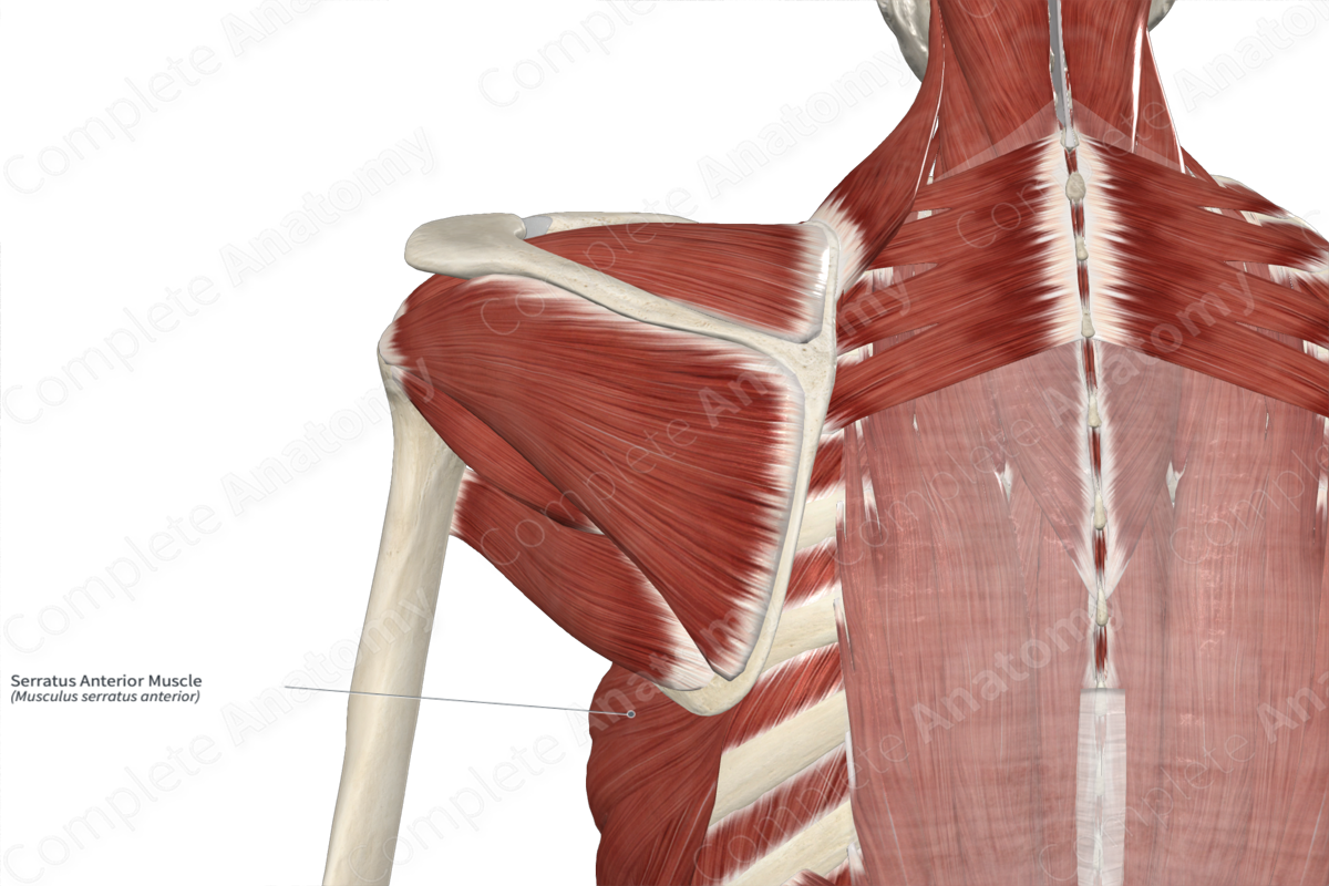
Insertion: Medial border of scapula.
Action: Protracts and upwardly rotates pectoral (shoulder) girdle at acromioclavicular and sternoclavicular joints.
Innervation: Long thoracic nerve (C5-C7).
Arterial Supply: Superior and lateral thoracic, thoracodorsal arteries.
Related parts of the anatomy
Origin
The serratus anterior muscle originates from the:
- external surfaces of the first to ninth ribs;
- fascia covering the external intercostal muscles.
There can be variations between individuals regarding the numbers of origin sites and the ribs to which they attach, with some only having origins on the first eight ribs, and others having origins on the first ten ribs (Standring, 2016).
Insertion
The fibers of the serratus anterior muscle travel posteriorly and medially around the thoracic cage and insert onto the entire length of the medial border of the scapula.
Key Features & Anatomical Relations
The serratus anterior muscle is found on the lateral and posterior aspects of the thorax. It is a broad skeletal muscle, and its digitations, found at its origin sites, give the muscle its saw-toothed or serrated appearance. It is located:
- superficial to the first eight ribs and the external intercostal muscles;
- deep to the scapula, the latissimus dorsi, trapezius, and subscapularis muscles, and the long thoracic nerve;
- posterior to the pectoralis major and minor muscles.
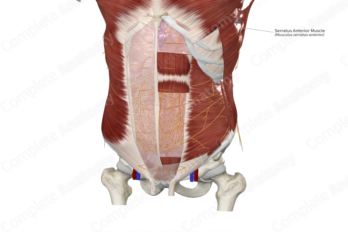
The serratus anterior muscle contributes to the formation of the medial wall of the axilla. At their origin sites, the lower fibers of the serratus anterior muscle interdigitate with the upper fibers of the external abdominal oblique muscle.
Actions & Testing
The serratus anterior muscle is involved in multiple actions:
- protracts the pectoral (shoulder) girdle at the acromioclavicular and sternoclavicular joints;
- upwardly rotates the pectoral girdle at the acromioclavicular and sternoclavicular joints;
- helps stabilize the scapula against the thoracic wall (Standring, 2016).
Because the serratus anterior muscle is used when punching, it is also known as “boxer’s muscle.” It can be tested by holding the shoulder in the flexed position and the elbow in the extending position, then pushing the upper limb against resistance, during which it can be seen and palpated. In cases of where this muscle is weak or paralyzed, the medial border of the scapula will become prominent on the upper back, a sign known as “winged scapula” (Sinnatamby, 2011).
List of Clinical Correlates
- Winged scapula
References
Moore, K. L., Dalley, A. F. and Agur, A. M. R. (2009) Clinically Oriented Anatomy. Lippincott Williams & Wilkins.
Sinnatamby, C. S. (2011) Last's Anatomy: Regional and Applied. ClinicalKey 2012: Churchill Livingstone/Elsevier.
Standring, S. (2016) Gray's Anatomy: The Anatomical Basis of Clinical Practice. Gray's Anatomy Series 41st edn.: Elsevier Limited.
Learn more about this topic from other Elsevier products

