Quick Facts
Origin: Clavicular head: anterior aspects of medial half of clavicle; Sternocostal head: manubrium, sternal body, the true ribs and their costal cartilages; Abdominal part: external abdominal oblique aponeurosis.
Insertion: Crest of greater tubercle of humerus.
Action: Adducts, medially rotates, and transversely adducts arm at glenohumeral (shoulder) joint; depresses pectoral (shoulder) girdle at acromioclavicular and sternoclavicular joints.
Innervation: Medial and lateral pectoral nerves (C5-T1).
Arterial Supply: Pectoral branches of thoracoacromial artery, perforating branches of internal thoracic artery, superior and lateral thoracic arteries.
Related parts of the anatomy
Origin
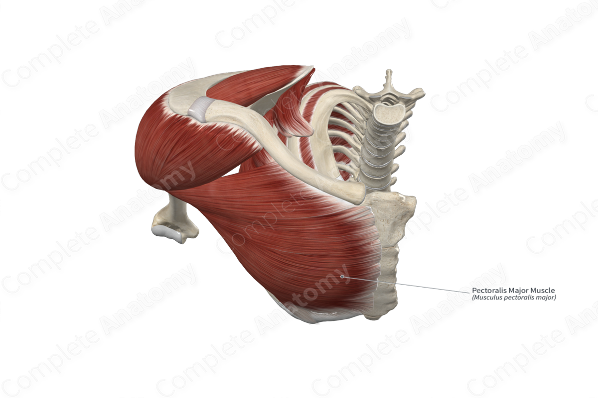
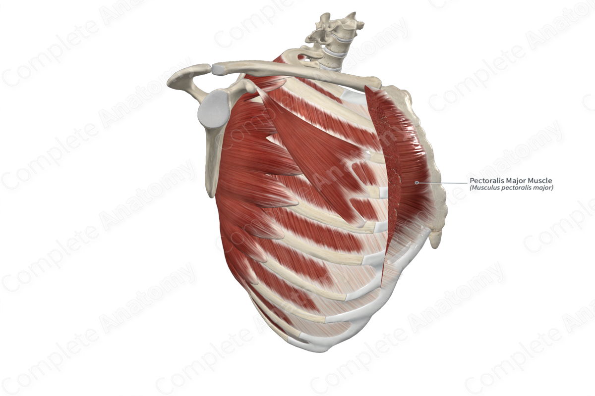
The pectoralis major muscle consists of three parts: the clavicular and sternocostal heads, and a variable abdominal part.
The clavicular head originates from the anterior aspect of the medial half of the clavicle.
The sternocostal head is the largest part and originates from the anterior aspects of the manubrium and body of the sternum, as well as from the anterior aspects of the superior six costal cartilages.
The abdominal part arises from the aponeurosis of the external abdominal oblique muscle; however, this head may be absent.
Additionally, the right and left pectoralis major muscles may insert into each other across the anterior sternal body.
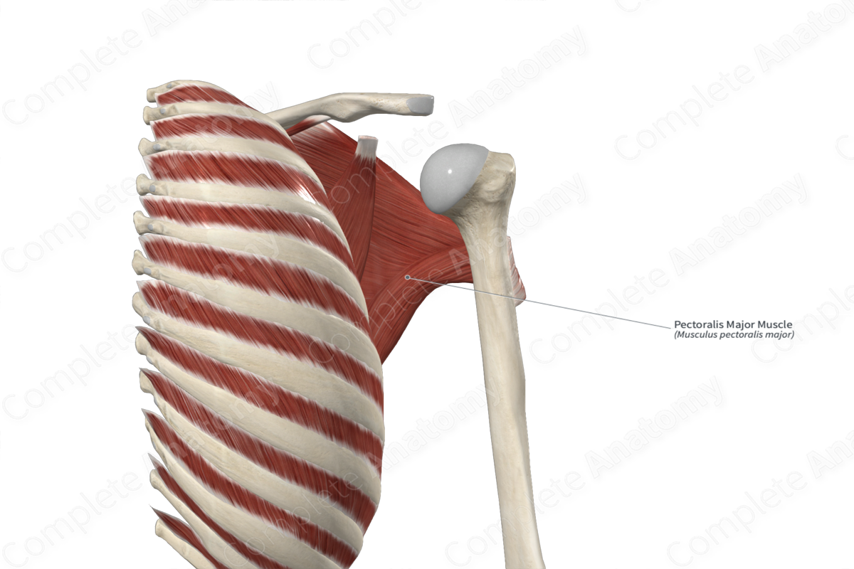
Insertion
The muscle bellies of the clavicular and sternocostal heads of pectoralis major travel laterally and converge to a single flat tendon, which inserts onto the crest of the greater tubercle of the humerus. This tendon consists of two laminae:
the anterior lamina, which is attached to the fibers that form the superior part of the muscle;
the posterior lamina, which is attached to the fibers that form the inferior part of the muscle.
The posterior lamina inserts more superiorly onto the humerus than the anterior lamina.
Key Features & Anatomical Relations
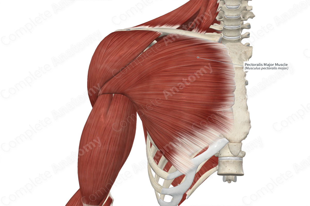
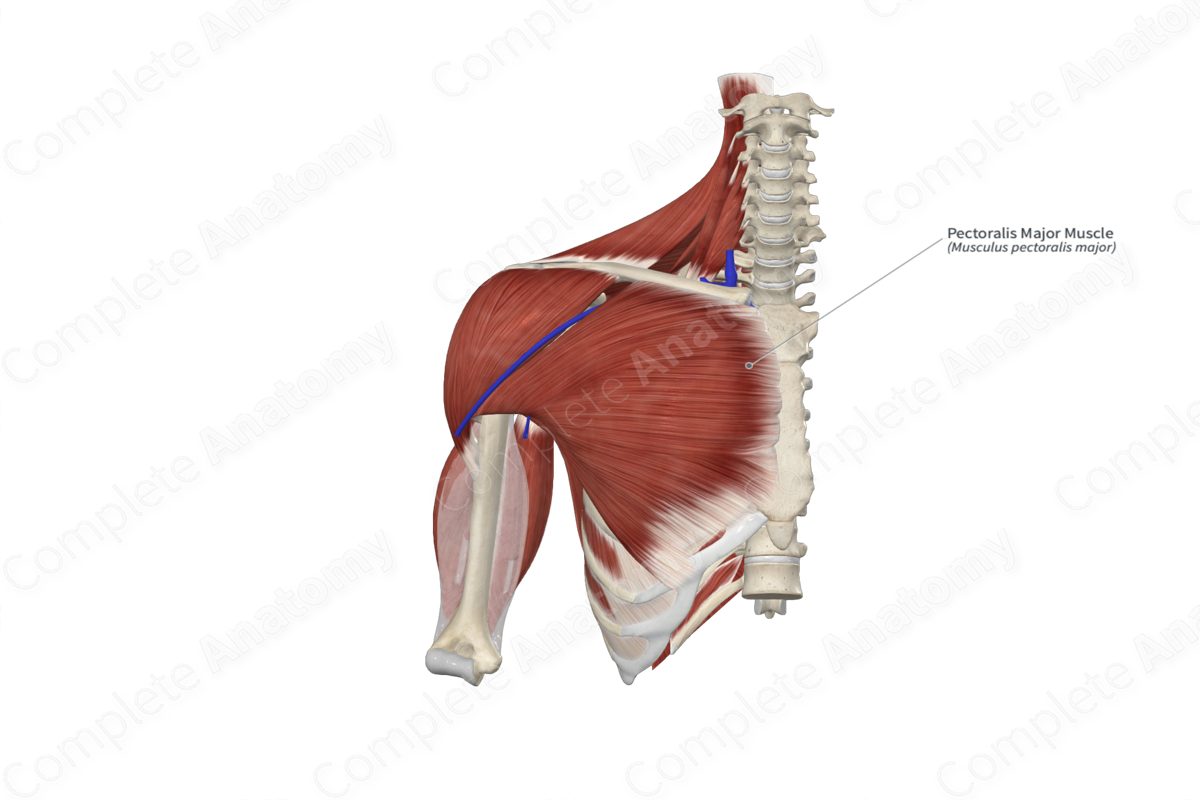
The pectoralis major muscle is found in the pectoral region of the thorax. It is a large, fan-shaped, convergent type of skeletal muscle. It is located:
anterior (superficial) to the sternum, the first six ribs and their costal cartilages, the pectoralis minor, serratus anterior, subclavius, and external intercostal muscles, and the external intercostal membrane;
posterior (deep) to the pectoral fascia and breast tissue;
inferomedial to the clavicular part of deltoid muscle.
The pectoralis major muscle contributes to the formation of the anterior wall of the axilla, and its inferior margin forms the anterior axillary fold. The space located between the superolateral margin of the pectoralis major muscle and the inferomedial margin of the clavicular part of deltoid muscle is known as the infraclavicular fossa, through which the cephalic vein passes.
Actions & Testing
The pectoralis major muscle is involved in multiple actions:
adducts the arm at the glenohumeral (shoulder) joint;
medially rotates the arm at the glenohumeral joint;
depresses the pectoral (shoulder) girdle at the acromioclavicular and sternoclavicular joints;
transversely adducts the arm at the glenohumeral joint (i.e., it adducts the flexed arm along the transverse plane) (via its clavicular head);
assists in flexion of the arm at the glenohumeral joint (via its clavicular head);
assists in extension of the arm at the glenohumeral joint (via its sternocostal head);
acts as an accessory muscle of inspiration.
The two heads of pectoralis major muscle can be tested separately.
The clavicular head can be tested by transversely adducting the arm at the glenohumeral joint against resistance, during which it can be seen and palpated.
The sternocostal head can be tested by adducting the arm at the glenohumeral joint against resistance, during which it can be seen and palpated (Standring, 2016).
List of Clinical Correlates
Poland’s syndrome
Chondro-epitrochlearis
References
Standring, S. (2016) Gray's Anatomy: The Anatomical Basis of Clinical Practice. Gray's Anatomy Series 41st edn.: Elsevier Limited.
Learn more about this topic from other Elsevier products

