Quick Facts
Origin: Lateral epicondyle of humerus, via common extensor tendon.
Insertion: Dorsal aspects of bases of both the middle and distal phalanges of the index, middle, ring, and little fingers.
Action: Extends index, middle, ring, and little fingers.
Innervation: Posterior antebrachial interosseous nerve (C7-C8).
Arterial Supply: Radial recurrent, anterior and posterior interosseous arteries.
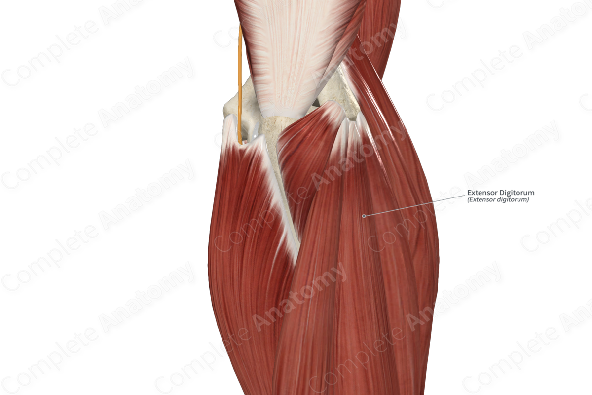
Origin
The extensor digitorum muscle originates from the:
- lateral epicondyle of humerus, via the common extensor tendon;
- adjacent antebrachial fascia.
The common extensor tendon is the combined tendon for four of the six superficial muscles in the posterior compartment of the forearm, which are the:
- extensor carpi radialis brevis muscle;
- extensor digitorum muscle;
- extensor digiti minimi muscle;
- extensor carpi ulnaris muscle.
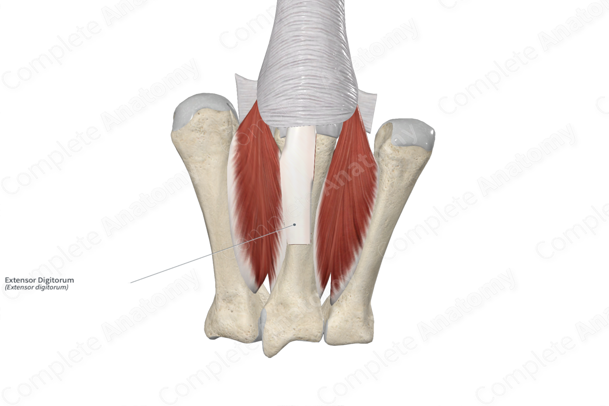
Insertion
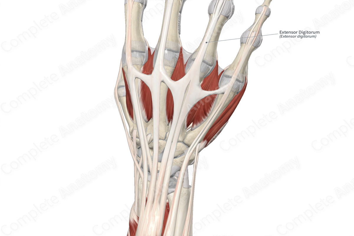
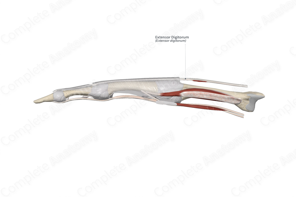
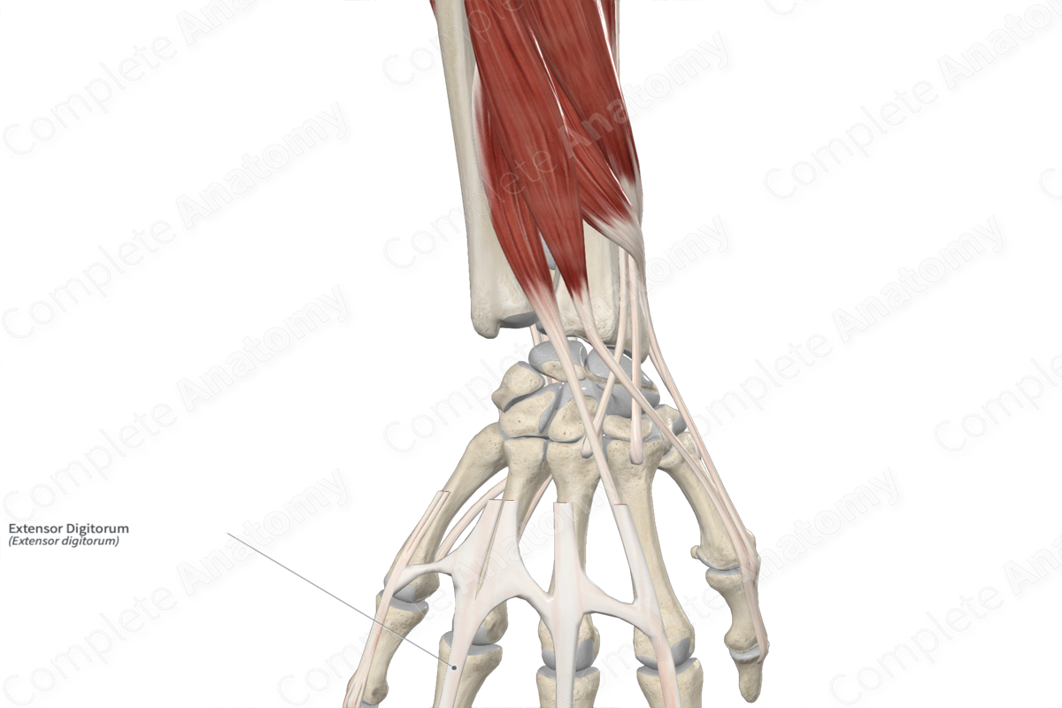
The fibers of the extensor digitorum muscle travel inferiorly to the hand and insert onto the:
- dorsal aspects of the bases of middle phalanges of the index, middle, ring, and little fingers;
- dorsal aspects of the bases of distal phalanges of the index, middle, ring, and little fingers.
Key Features & Anatomical Relations
The extensor digitorum muscle is one of the muscles of the superficial part of the posterior compartment of the forearm. It is a long, broad, fusiform type of skeletal muscle that divides into four tendons in the distal forearm.
At the wrist, its tendons travel deep to the extensor retinaculum of hand, where they pass through the common tendinous sheath of extensor digitorum and extensor indicis. Along the dorsal aspect of the hand, the tendons of the extensor digitorum spread out as they travel to their respective fingers, with intertendinous connections linking them together. Along the dorsal aspects of the phalanges of the index, middle, ring and little fingers, the tendons each form an aponeurotic extensor expansion, which then travel to their insertion sites.
The extensor digitorum muscle is located:
- Posterior (superficial) to the supinator, abductor pollicis longus, extensor pollicis longus, and extensor indicis muscles;
- Medial to the extensor carpi radialis brevis muscle.
- Lateral to the extensor digiti minimi and extensor carpi ulnaris muscles.
Actions & Testing
The extensor digitorum muscle is involved in multiple actions:
- extends the distal phalanges at the distal interphalangeal joints of the index, middle, ring, and little fingers;
- extends the middle phalanges at the proximal interphalangeal joints of the same fingers;
- extends the proximal phalanges at the metacarpophalangeal joints of the same fingers;
- assists in extension of the hand at the radiocarpal (wrist) joint.
The extensor digitorum muscle can be tested by extending the fingers against resistance while the forearm is held in the pronated position, during which it can be seen and palpated (Standring, 2016).
List of Clinical Correlates
- Lateral epicondylitis (tennis elbow)
References
Standring, S. (2016) Gray's Anatomy: The Anatomical Basis of Clinical Practice. Gray's Anatomy Series 41st edn.: Elsevier Limited.
Learn more about this topic from other Elsevier products

