Quick Facts
Origin: Lateral epicondyle of humerus, via common extensor tendon.
Insertion: Extensor expansion of little finger.
Action: Extends little finger.
Innervation: Posterior antebrachial interosseous nerve (C7-C8).
Arterial Supply: Radial recurrent, anterior and posterior interosseous arteries.


Origin
The extensor digiti minimi muscle originates from the:
- lateral epicondyle of humerus, via the common extensor tendon;
- adjacent antebrachial fascia.
The common extensor tendon is the combined tendon for four of the six superficial muscles in the posterior compartment of the forearm, which are the:
- extensor carpi radialis brevis muscle;
- extensor digitorum muscle;
- extensor digiti minimi muscle;
- extensor carpi ulnaris muscle.
Insertion
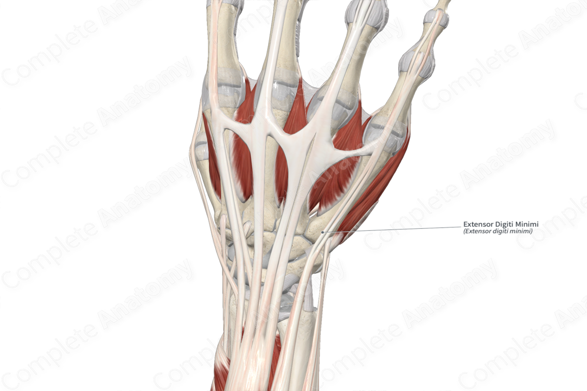
The fibers of the extensor digiti minimi muscle travel inferomedially to the hand and insert, via a long tendon, onto the extensor expansion of the tendon of extensor digitorum muscle that goes to the little finger.
Key Features & Anatomical Relations
The extensor digiti minimi muscle is one of the muscles of the superficial part of the posterior compartment of the forearm. It is a thin, long, fusiform type of skeletal muscle.
From its origin, its muscle belly fibers can appear blended with those of the extensor digitorum muscle; however, it becomes a distinct muscle in the middle of the forearm. In the distal one third of the forearm, the muscle belly gives rise to a long tendon, which travels inferomedially along the posterior aspect of the forearm. At the wrist, its tendon travels deep to the extensor retinaculum of hand, where it passes through the tendinous sheath of extensor digiti minimi. Within the hand, the tendon then travels inferomedially to its insertion site.
The extensor digiti minimi muscle is located:
- posterior (superficial) to the ulna and the proximal ends of the supinator, abductor pollicis longus, extensor pollicis longus, and extensor indicis muscles;
- medial to the extensor digitorum muscle;
- lateral to the extensor carpi ulnaris muscle.
Actions & Testing
The extensor digiti minimi muscle is involved in multiple actions:
- extends the distal phalanx of little finger at its distal interphalangeal joint;
- extends the middle phalanx of little finger at its proximal interphalangeal joint;
- extends the proximal phalanx of little finger at its metacarpophalangeal joint;
- assists in extension of the hand at the radiocarpal (wrist) joint.
This muscle can contract with the extensor digitorum during extension of all four fingers, or contract alone to fully extend the little finger while the other fingers remain in their fully flexed positions.
The extensor digiti minimi muscle can be tested by extending the index finger while the other three fingers are held in the flexed position. This position of the other three fingers prevents the extensor digitorum muscle from contracting (Standring, 2016).
List of Clinical Correlates
- Lateral epicondylitis (tennis elbow)
References
Standring, S. (2016) Gray's Anatomy: The Anatomical Basis of Clinical Practice. Gray's Anatomy Series 41st edn.: Elsevier Limited.
Learn more about this topic from other Elsevier products

