Quick Facts
Origin: Xiphoid process, internal surfaces of seventh to twelfth costal cartilages and adjacent ribs, medial and lateral arcuate ligaments, L1-L3 vertebrae.
Insertion: Central tendon.
Action: Primary muscle of inspiration.
Innervation: Motor: phrenic nerves (C3-C5); Sensory: phrenic nerves and lower intercostal nerves.
Arterial Supply: Superior and inferior phrenic, pericardiacophrenic, and musculophrenic arteries.
Origin
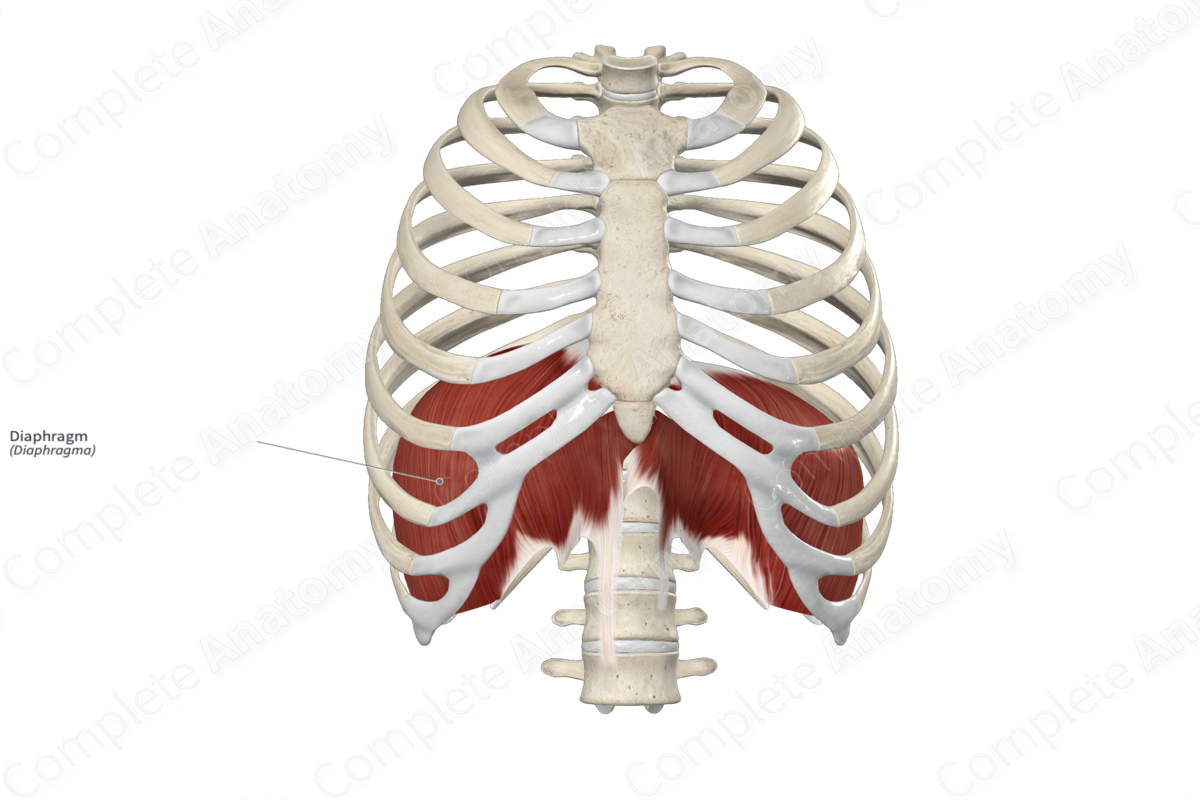
The fibers of the diaphragm can be divided into sternal, costal, and lumbar parts.
- The sternal part originates from the posterior aspect of the xiphoid process.
- The costal part originates from the internal surfaces of the seventh to twelfth costal cartilages and adjacent ribs on both the right and left sides.
- The lumbar part originates from the medial and lateral arcuate ligaments and, via its left and right crura, and from the first to third lumbar vertebrae.
Insertion
The fibers of the sternal, costal, and lumbar parts of the diaphragm converge and insert onto a single aponeurosis known as the central tendon.
Key Features & Anatomical Relations
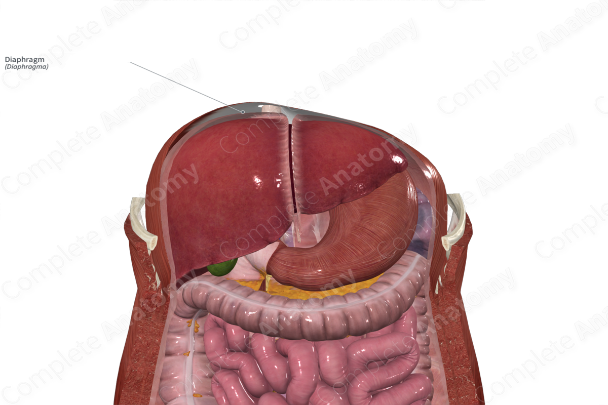
For classification purposes, the diaphragm is also considered one of the muscles of the thorax. It is a broad, double-domed, fibromuscular structure that separates the thoracic cavity above from the abdominal cavity below.
The diaphragm consists of right and left muscular domes and a fibrous central tendon. Its muscular fibers can be divided into the:
- anteriorly located sternal part;
- laterally located costal part;
- posteriorly located lumbar part.
The lumbar part consists of two fibromuscular bands, the right and left crura, whose fibers blend with the anterior longitudinal ligament. The right crus is long and attaches to the vertebral bodies of the first to third lumbar vertebrae. It also gives off fibers that contribute to the formation of the suspensory ligament of duodenum. The left crus is shorter and attaches to the vertebral bodies of the first to second lumbar vertebrae.
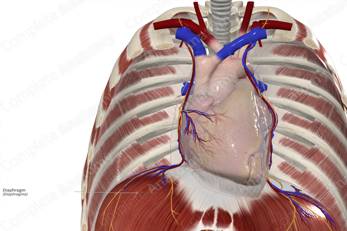
The central tendon is the broad, thin, trifoliate aponeurosis that is located close to the center of the diaphragm. Its fibers blend with the fibrous pericardium superiorly.
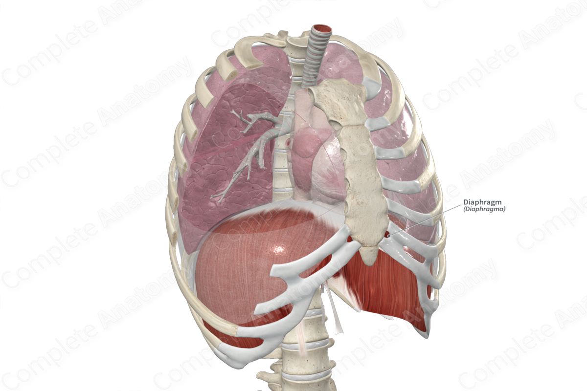
The diaphragm is located:
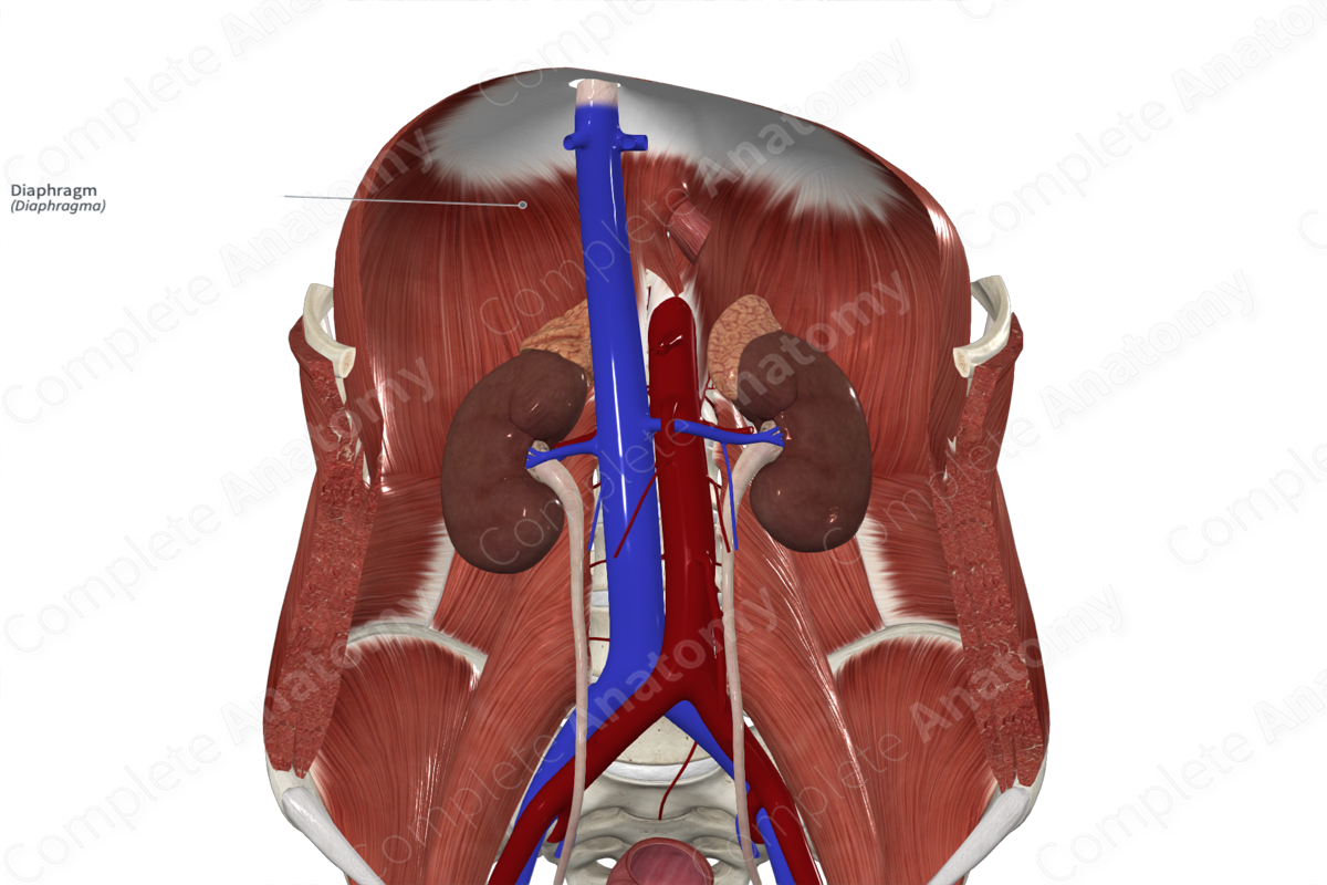
- superior to the peritoneum, liver, kidneys, suprarenal glands, stomach and spleen;
- inferior to the lungs, the pleurae, and the heart and its pericardium;
The right muscular dome of the diaphragm is higher than the left, this is due to the position of the right lobe of the liver.
In order to allow for the passage of structures between the thorax and abdomen, the diaphragm has apertures:
- the caval foramen (for the inferior vena cava) is present in the central tendon, it lies at the level of the eighth thoracic vertebra (T8);
- the esophageal hiatus (for the esophagus) is present along the right crus, it lies at the level of the tenth thoracic vertebra (T10);
- the aortic hiatus (for the aorta) is present posterior to the median arcuate ligament, it lies at the level of the twelfth thoracic vertebra (T12).
Actions
The diaphragm is the primary muscle of inspiration. During contraction, it descends, increasing thoracic volume and decreasing thoracic pressure, resulting in inspiration. Conversely, during relaxation, it ascends, decreasing thoracic volume and increasing thoracic pressure, resulting in quiet passive expiration (Sinnatamby, 2011).
List of Clinical Correlates
- Hiatus hernia
- Congenital diaphragmatic hernia
- Paralysis of diaphragm
References
Sinnatamby, C. S. (2011) Last's Anatomy: Regional and Applied. ClinicalKey 2012: Churchill Livingstone/Elsevier.
Learn more about this topic from other Elsevier products

