Quick Facts
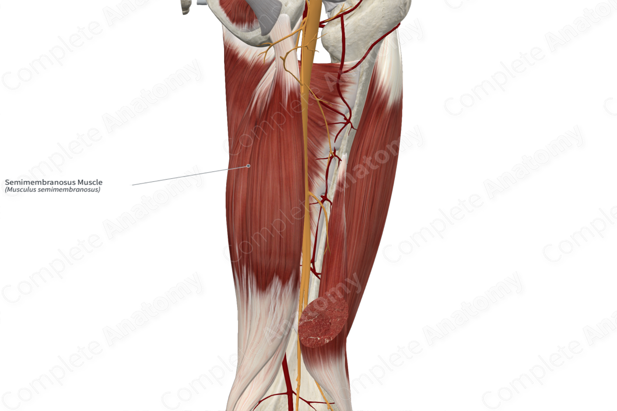
Origin: Ischial tuberosity.
Insertion: Medial condyle of tibia.
Action: Flexes and medially rotates leg at knee joint; extends thigh at hip joint.
Innervation: Tibial division of sciatic nerve (L5-S2).
Arterial Supply: Perforating arteries of deep femoral artery.
Related parts of the anatomy
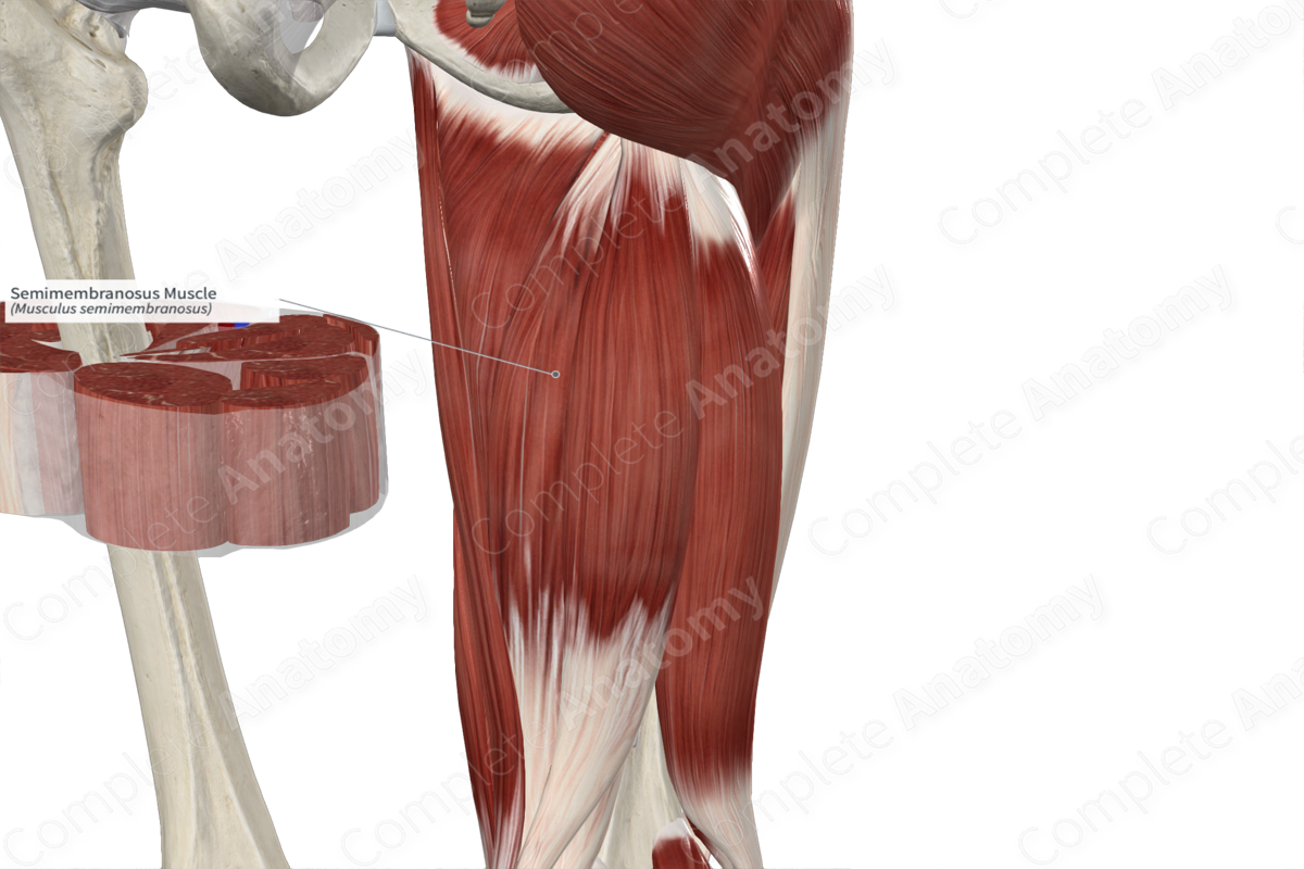
Origin
The semimembranosus muscle originates from the lateral aspect of the posterior portion of the ischial tuberosity. The proximal tendon of the semimembranosus is long and flat, with a membranous appearance.
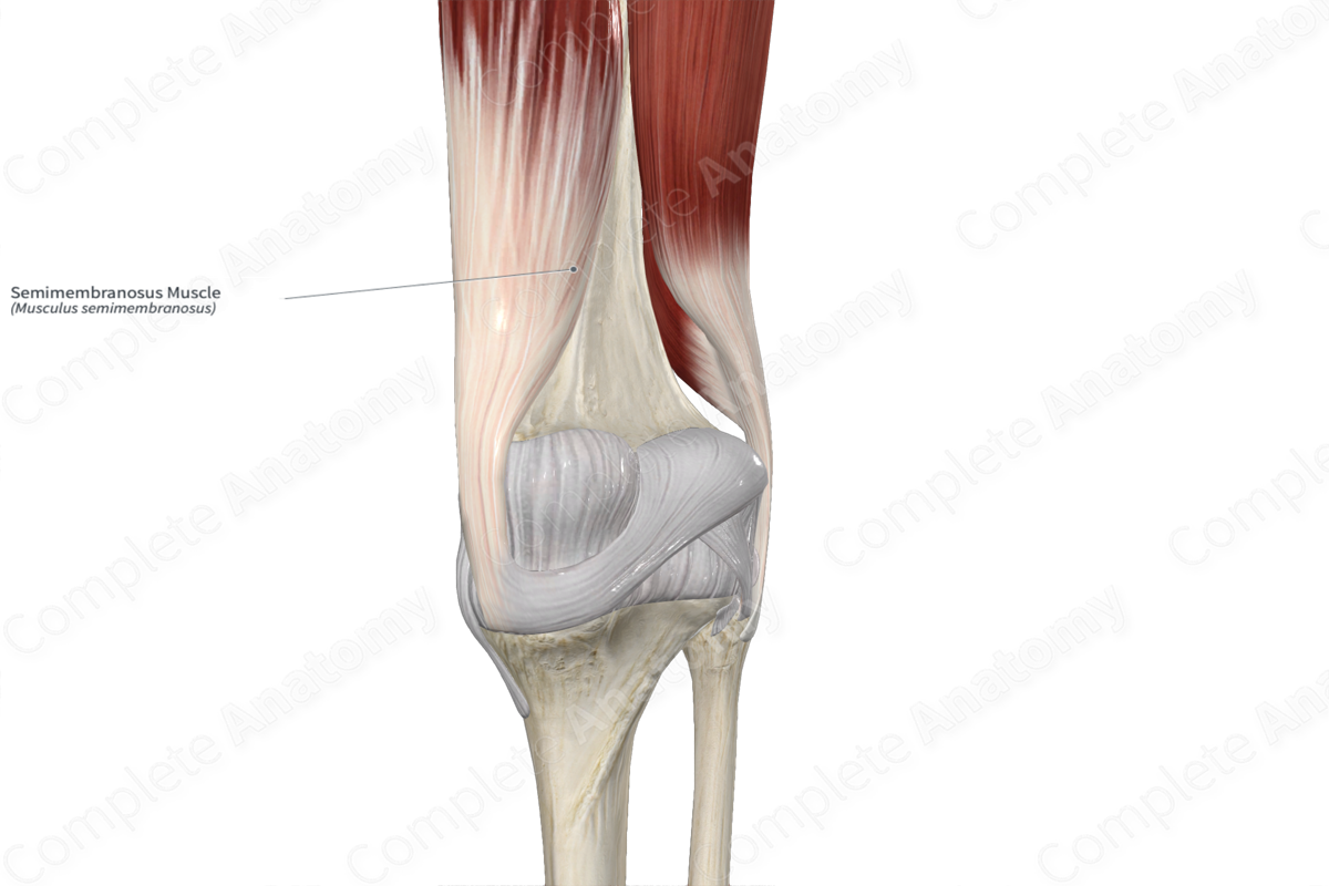
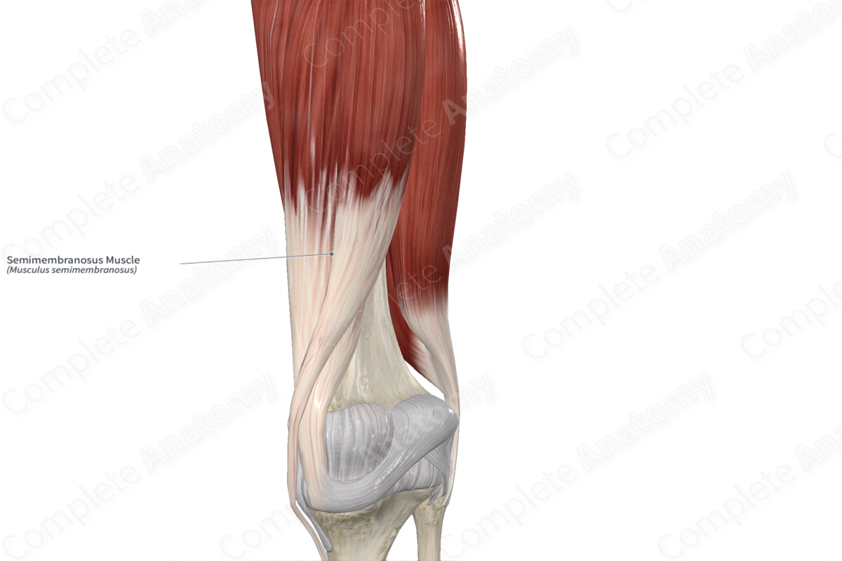
Insertion
The fibers of the semimembranosus muscle travel inferiorly and insert, via a rounded tendon, onto the posterolateral aspect of the medial condyle of the tibia. From the distal tendon of the semimembranosus arises the oblique popliteal ligament.
Key Features & Anatomical Relations
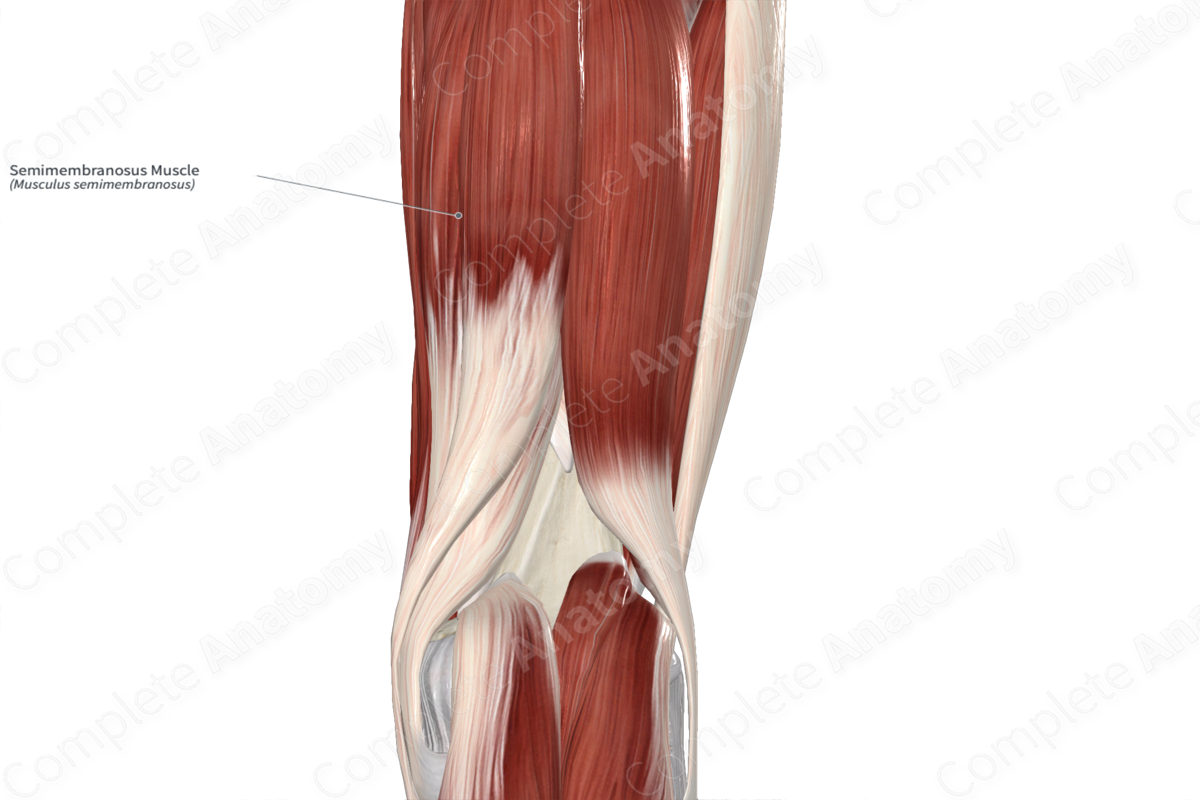
The semimembranosus muscle is found in the posterior compartment of the thigh. It is a long, unipennate type of skeletal muscle.
It is located:
- anterior to the semitendinosus muscle;
- posterior to the adductor magnus muscle;
- medial to the long head of biceps femoris muscle, the sciatic nerve, and the semimembranosus bursa;
- lateral to the gracilis muscle (at its distal end).
The semimembranosus muscle contributes to the formation of the popliteal fossa, where the muscle and tendon form its superomedial boundary.
The term “hamstrings” is the collective name given to the long head of biceps femoris, semitendinosus, and semimembranosus muscles. These three muscles share similar features that the short head of biceps femoris does not, including:
- originating from the ischial tuberosity;
- acting on both the hip and knee joints;
- innervated by the tibial division of sciatic nerve.
Actions & Testing
The semimembranosus muscle is involved in multiple actions:
- flexes the leg at the knee joint;
- medially rotates the leg at the knee joint while this joint is held in a semiflexed position;
- extends the thigh at the hip joint.
The semimembranosus muscle cannot be tested in isolation, therefore all of the muscles of the posterior compartment of the thigh are tested simultaneously by flexing the leg at the knee joint against resistance while lying in the prone position, during which the semimembranosus tendon can be palpated (Standring, 2016).
List of Clinical Correlates
- Hamstring strain
- Hamstring tear
- Avulsion of ischial tuberosity
References
Standring, S. (2016) Gray's Anatomy: The Anatomical Basis of Clinical Practice. Gray's Anatomy Series 41st edn.: Elsevier Limited.
Learn more about this topic from other Elsevier products

