Structure/Morphology
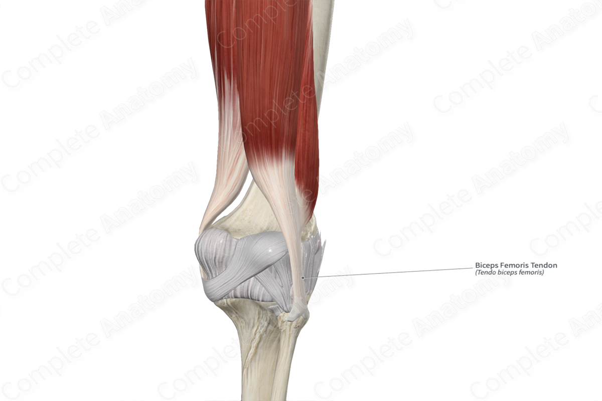
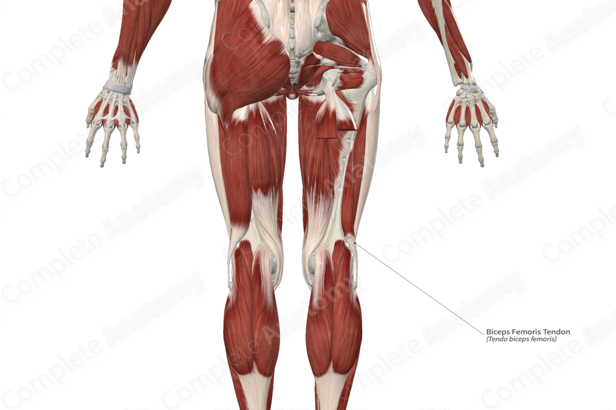
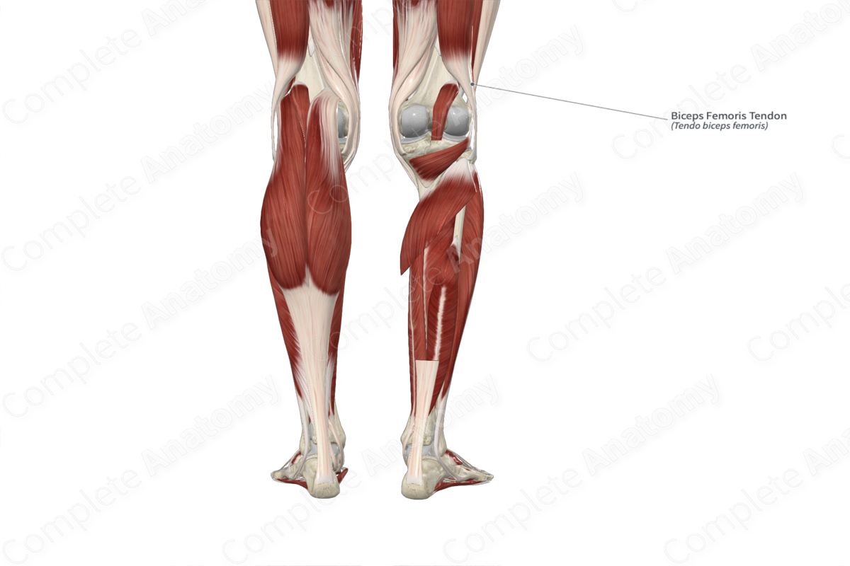
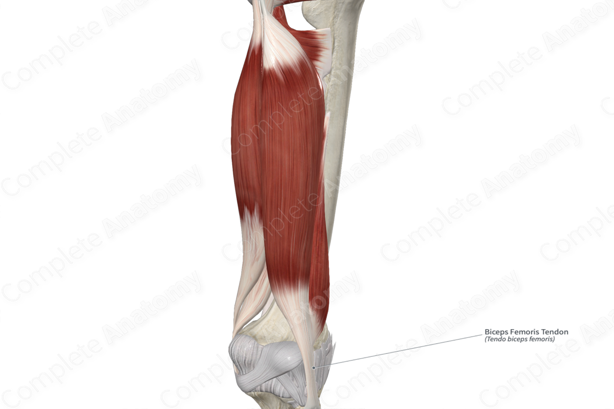
The biceps femoris tendon is the rounded tendon that attaches the muscle bellies of the long and short heads of the biceps femoris muscle to the head of the fibula. The proximal end of the tendon begins in the distal one third of the thigh, where the fibers of the two heads of the biceps femoris muscle converge to form this single tendon. The distal end of the biceps femoris tendon splits around the fibular collateral ligament and then attaches to the lateral aspect of the head of the fibula.
Related parts of the anatomy
Anatomical Relations
The biceps femoris tendon is located:
- anterior to the lateral head of gastrocnemius muscle and the common fibular nerve;
- lateral to the lateral condyle of femur, capsule of the knee joint, the fibular collateral ligament, and the inferior subtendinous bursa of biceps femoris muscle.
Overall, the biceps femoris muscle contributes to the formation of the popliteal fossa, where the muscle and tendon form its superolateral boundary.
Function
The biceps femoris tendon attaches the muscle bellies of the long and short heads of the biceps femoris muscle to the head of the fibula.
This insertion site allows the biceps femoris muscle to be able to:
- flex the leg at the knee joint;
- laterally rotate the leg at the knee joint while this joint is held in a semiflexed position.
List of Clinical Correlates
- Biceps femoris tendinitis
Learn more about this topic from other Elsevier products

