
Quick Facts
Origin: Pelvic surface of sacrum and sacrotuberous ligament.
Insertion: Superior border of greater trochanter of femur.
Action: Laterally rotates and transversely abducts thigh at hip joint.
Innervation: Nerve to piriformis (S1-S2).
Arterial Supply: Superior and inferior gluteal, internal pudendal, and lateral sacral arteries.


Related parts of the anatomy
Origin
The piriformis muscle originates from the:
- area of the pelvic surface of the sacrum that is located between the second, third, and fourth anterior sacral foramina;
- sacrotuberous ligament.
Insertion
The fibers of the piriformis muscle travel anterolaterally and insert, via a rounded tendon, onto the superior border of the greater trochanter of the femur.
Key Features & Anatomical Relations
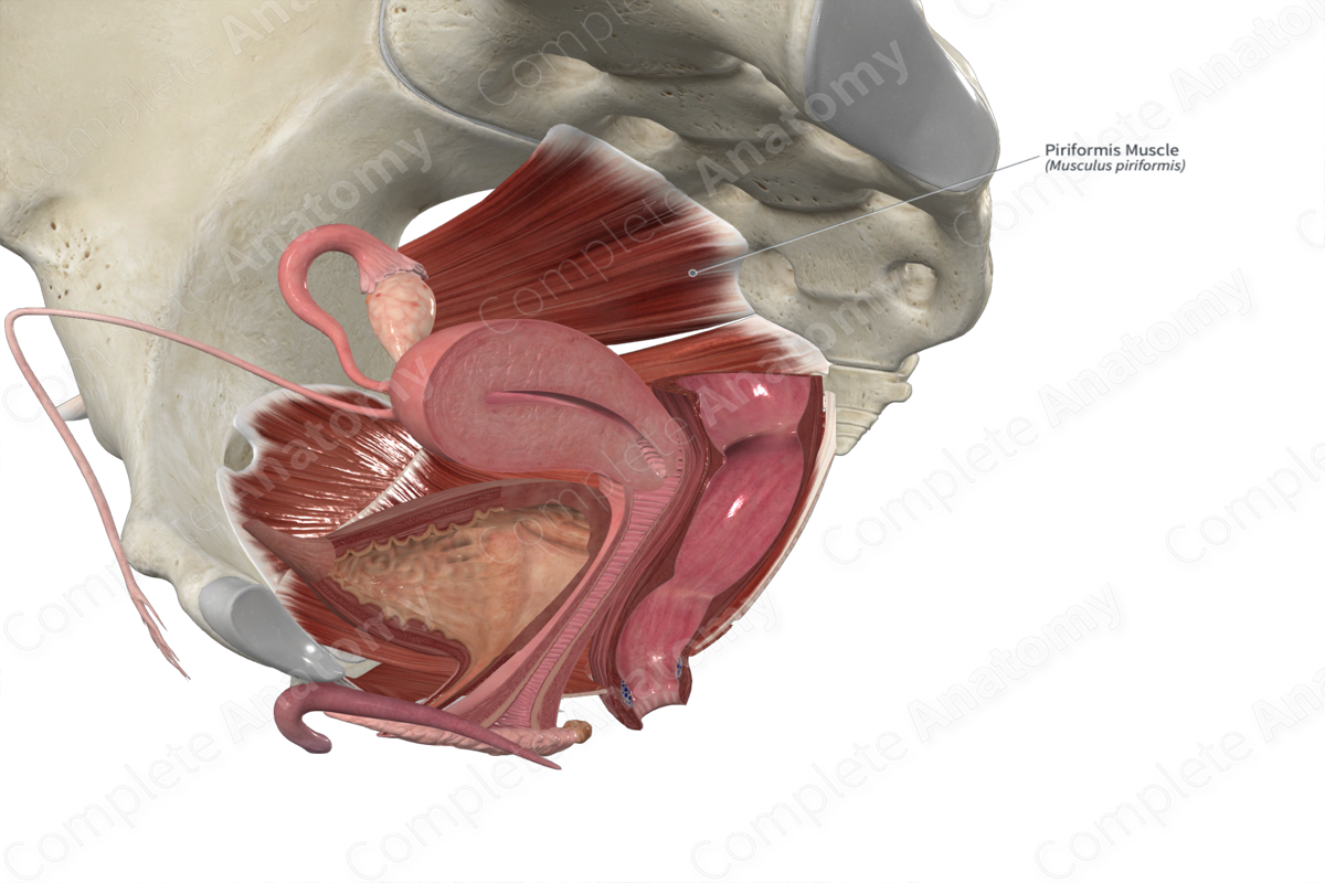
The piriformis muscle is one of the deep gluteal muscles. It is a thick, pear-shaped, convergent type of skeletal muscle. Within the lesser pelvis, the fibers of the piriformis muscle travel anterolaterally to the greater sciatic foramen. After passing through the foramen, it enters the gluteal region and travels posterior to the hip joint, where its muscle belly gives rise to a rounded tendon that travels inferolaterally to its insertion site.
The piriformis muscle is located:
- anterior to the sacrum and the gluteus maximus muscle;
- posterior to the capsule of the hip joint, the sacral plexus, and the sciatic nerve;
- superior to the coccygeus and superior gemellus muscles, and the inferior gluteal artery and nerve;
- inferior to the gluteus medius muscle, and the superior gluteal artery and nerve.
Actions & Testing
The piriformis muscle is involved in multiple actions:
- laterally rotates the thigh at the hip joint;
- transversely abducts the thigh at the hip joint (i.e., it abducts the flexed thigh along the transverse plane).
The piriformis muscle cannot be tested in isolation; therefore it is tested simultaneously with the obturator externus and quadratus femoris muscles and the three muscles of the triceps coxae (i.e., superior and inferior gemelli and obturator internus muscles) by laterally rotating the thigh at the hip joint against resistance (Standring, 2016).
List of Clinical Correlates
- Piriformis syndrome
References
Standring, S. (2016) Gray's Anatomy: The Anatomical Basis of Clinical Practice. Gray's Anatomy Series 41st edn.: Elsevier Limited.
Learn more about this topic from other Elsevier products

