Quick Facts
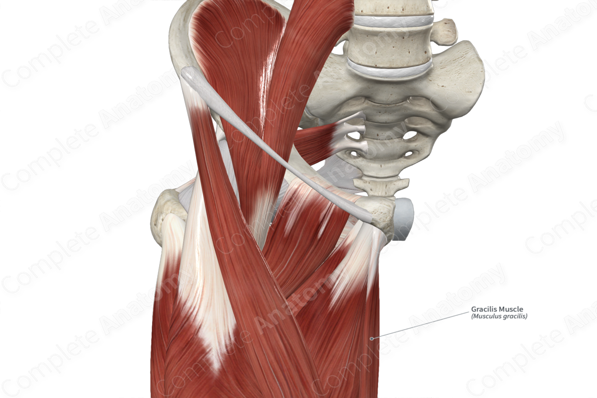
Origin: Body of pubis and inferior pubic ramus.
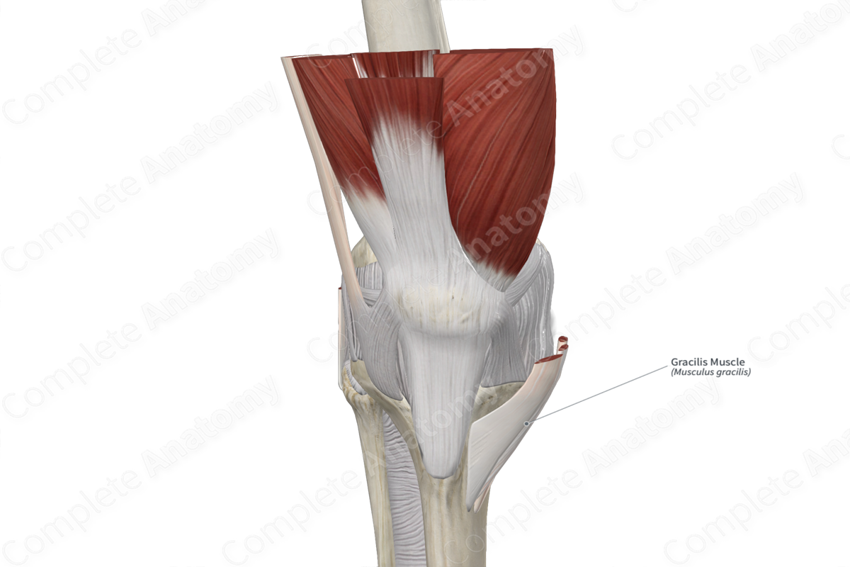
Insertion: Medial aspect of proximal part of tibia.
Action: Flexes and medially rotates leg at knee joint; assists in adduction of thigh at hip joint.
Innervation: Obturator nerve (L2-L3).
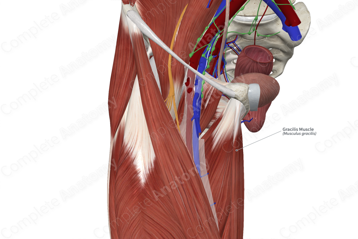
Arterial Supply: Deep femoral and medial circumflex femoral arteries.
Related parts of the anatomy
Origin
The gracilis muscle originates from the:
- anterior surface of the body of pubis;
- external surface of inferior pubic ramus.
Insertion
The fibers of the gracilis muscle travel inferiorly and insert, via a rounded tendon, onto the medial aspect of the proximal part of tibia.
Key Features & Anatomical Relations
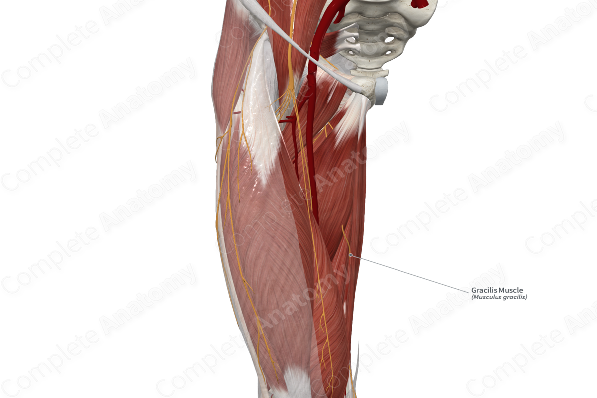
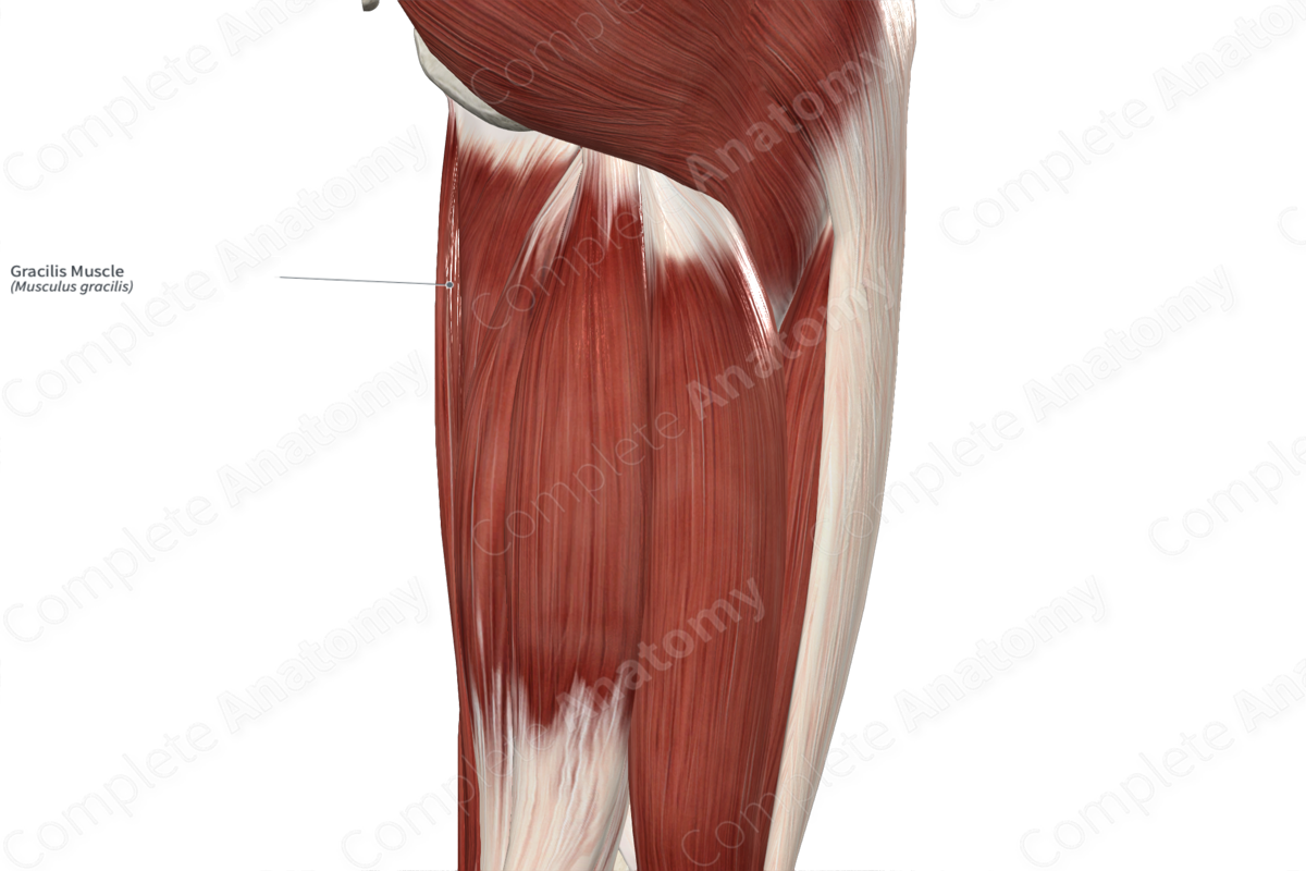
The gracilis muscle is found in the medial compartment of the thigh. It is a long, narrow, strap-like type of skeletal muscle. In the distal one third of the thigh, the muscle belly gives rise to a rounded tendon, which travels inferiorly. It passes medial to the knee joint, between the tendons of the sartorius and semitendinosus muscles, and then to its insertion site. At their insertion sites, the tendons of the gracilis, sartorius and semitendinosus muscles collectively form the pes anserinus, which is Latin for goose’s foot.
The gracilis muscle is located:
- anterior to the semitendinosus muscle (at its distal end);
- posterior to the sartorius muscle (at its distal end);
- medial to the adductor longus, adductor magnus and semimembranosus muscles, the tibial collateral ligament, and the anserine bursa.
Actions & Testing
The gracilis muscle is involved in multiple actions:
- medially rotates the leg at the knee joint while this joint is held in a semiflexed position;
- assists in flexion of the leg at the knee joint;
- assists in adduction of the thigh at the hip joint (Standring, 2016).
The gracilis muscle cannot be tested in isolation, therefore all of the muscles of the medial compartment of the thigh are tested simultaneously by adducting the thigh at the hip joint against resistance while lying in the supine position with the knee extended, during which the muscle can be palpated (Sinnatamby, 2011).
List of Clinical Correlates
- Gracilis musculocutaneous flap
References
Sinnatamby, C. S. (2011) Last's Anatomy: Regional and Applied. ClinicalKey 2012: Churchill Livingstone/Elsevier.
Standring, S. (2016) Gray's Anatomy: The Anatomical Basis of Clinical Practice. Gray's Anatomy Series 41st edn.: Elsevier Limited.
Learn more about this topic from other Elsevier products

