Quick Facts
Origin: Posterior surface of tibia, inferior to soleal line.
Insertion: Plantar aspects of bases of the distal phalanges of the second, third, fourth, and little toes
Action: Flexes second, third, fourth, and little toes; plantarflexes foot at ankle joint.
Innervation: Tibial nerve (L5-S2).
Arterial Supply: Posterior tibial artery.
Related parts of the anatomy
Origin
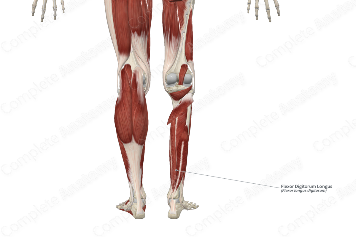
The flexor digitorum longus muscle originates from the medial aspect of the area of the posterior surface of the tibia that is located inferior to the soleal line.
Insertion
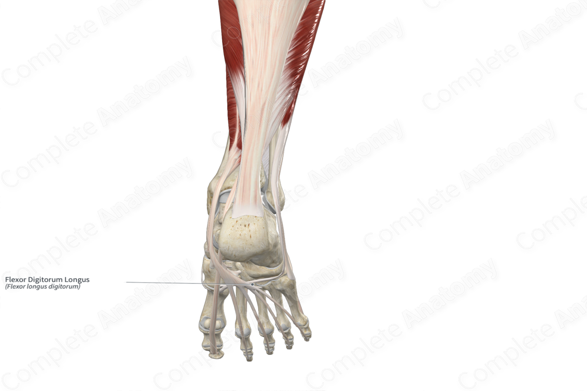
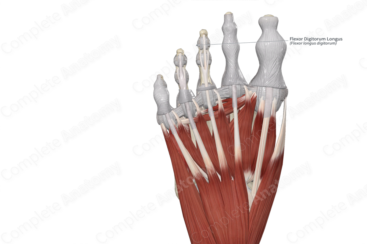
The fibers of the flexor digitorum longus muscle travel inferiorly to the foot and insert, via four separate tendons, onto the plantar aspects of the bases of distal phalanges of the second, third, fourth, and little toes.
Key Features & Anatomical Relations
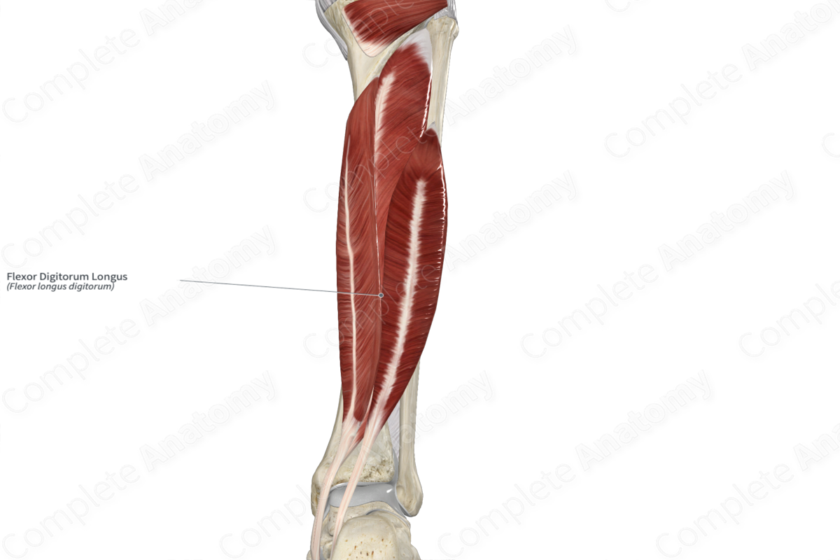
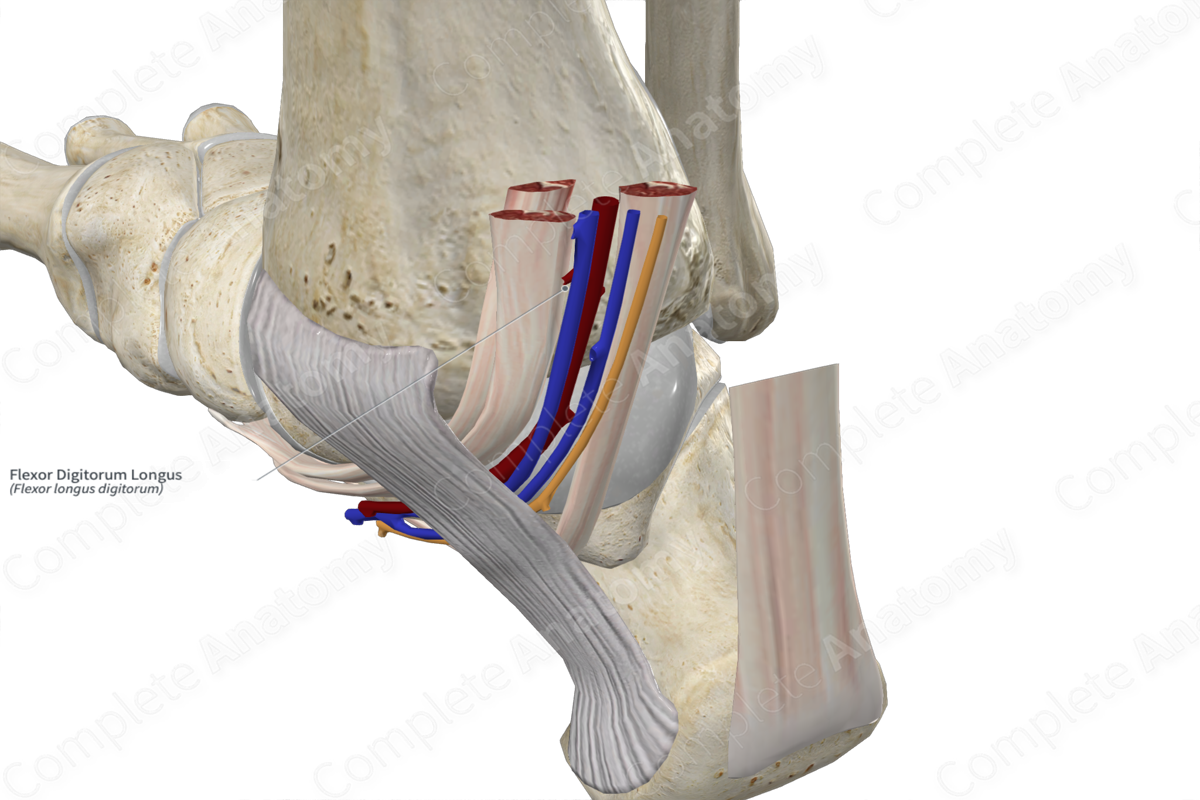
The flexor digitorum longus muscle is one of the muscles of the deep part of the posterior compartment of the leg. It is a long, narrow, bipennate type of skeletal muscle. In the distal one third of the leg, the muscle belly gives rise to a tendon that travels inferiorly towards the malleolar groove of the tibia. The tendon hooks around the groove, changing its line of pull to a more anteroinferior direction. This tendon then travels deep to the flexor retinaculum of foot, posterior to the adjacent tendon of tibialis posterior, where it passes through the tendinous sheath of flexor digitorum longus muscle.
Within the plantar part of the foot, the tendon travels anterolaterally and divides into four tendons. Each tendon travels deep to the tendons of the flexor digitorum brevis muscle. The tendons of both muscles travel through the fibrous sheaths that are found along the plantar aspects of the second, third, fourth and little toes. Along each proximal phalanx, the tendons of the flexor digitorum longus pass through the tendons of the flexor digitorum brevis, and then travel to their insertion sites.
Within the posterior compartment of the leg, the flexor digitorum longus muscle is located:
- anterior to the transverse intermuscular septum of leg and the soleus muscle;
- posterior to the tibia;
- medial to the flexor hallucis longus and tibialis posterior muscles, the posterior tibial vessels, and the tibial nerve.
Within the plantar part of the foot:
- the tendon of the flexor digitorum longus is located superficial to the tendon of the flexor hallucis longus and deep to the abductor hallucis and flexor digitorum brevis muscles;
- each of the four lumbrical muscles of the foot originates from each of the four tendons of the flexor digitorum longus;
- the quadratus plantae muscle inserts onto the tendons of the flexor digitorum longus.
The flexor digitorum longus muscle is one of five structures that pass posterior to the medial malleolus. From anterior to posterior, these are the tibialis posterior muscle, flexor digitorum longus muscle, posterior tibial artery, tibial nerve, and flexor hallucis longus muscle.
Actions & Testing
The flexor digitorum longus muscle is involved in multiple actions:
- flexes the distal phalanges at the distal interphalangeal joints of the second, third, fourth and little toes;
- flexes the middle phalanges at the proximal interphalangeal joints of the same toes;
- flexes the proximal phalanges at the metatarsophalangeal joints of the same toes;
- plantarflexes the foot at the ankle joint;
- helps stabilize the longitudinal arch of the foot (Sinnatamby, 2011).
The flexor digitorum longus muscle cannot be tested in isolation; therefore it is tested simultaneously with the flexor digitorum brevis muscle by flexing the second, third, fourth, and little toes against resistance, during which its tendons can be palpated (Standring, 2016).
List of Clinical Correlates
- Hammer toe deformities
References
Sinnatamby, C. S. (2011) Last's Anatomy: Regional and Applied. ClinicalKey 2012: Churchill Livingstone/Elsevier.
Standring, S. (2016) Gray's Anatomy: The Anatomical Basis of Clinical Practice. Gray's Anatomy Series 41st edn.: Elsevier Limited.
Learn more about this topic from other Elsevier products
Flexor Digitorum Longus Muscle

