Quick Facts
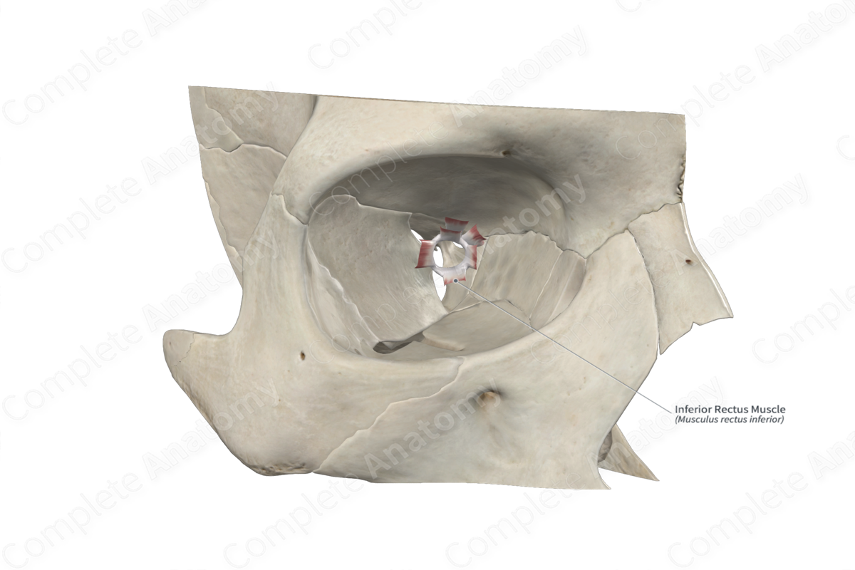
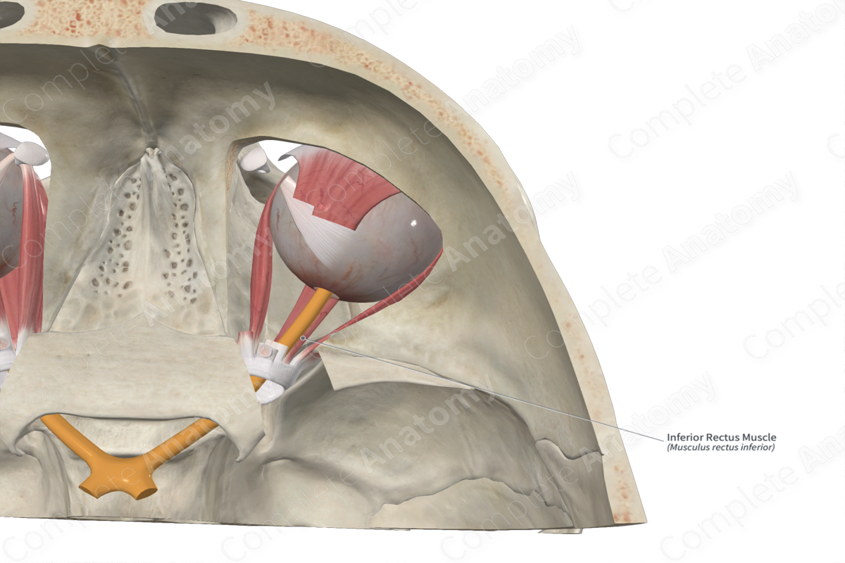
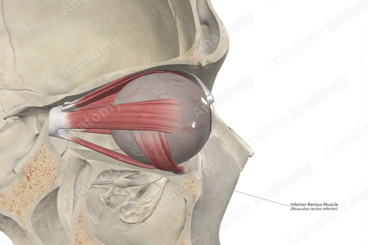
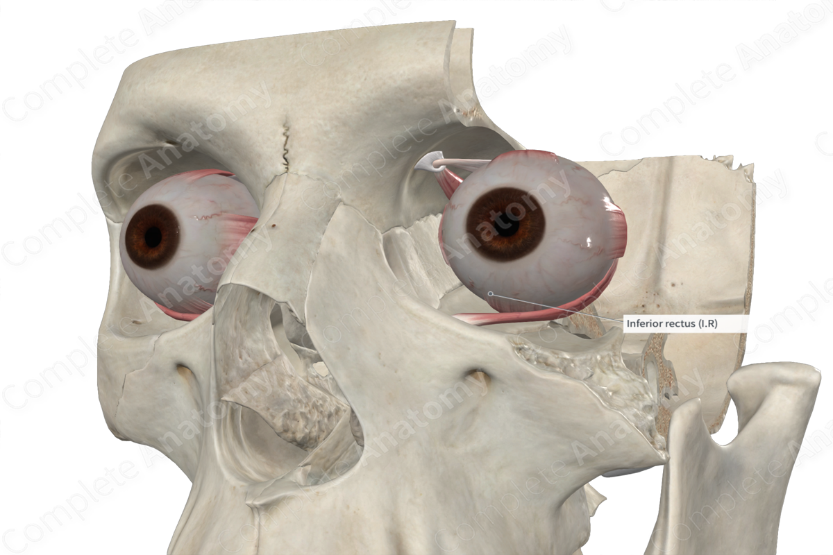
Origin: Common tendinous ring.
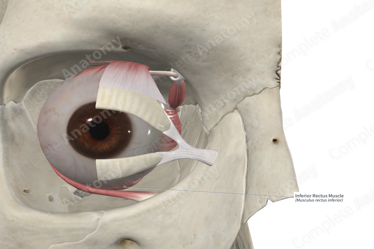
Insertion: Sclera, on inferior aspect of eyeball, just posterior to the corneoscleral junction.
Action: Depresses, adducts, and laterally rotates eyeball.
Innervation: Inferior branch of oculomotor nerve (CN III).
Arterial Supply: Infraorbital and ophthalmic arteries.
Origin
The inferior rectus muscle arises from the common tendinous ring, just below the optic canal.
Insertion
The inferior rectus muscle inserts obliquely on the sclera on the inferior aspect of the eyeball, posterior to the corneoscleral junction. Additional fibers extend from the inferior rectus muscle to the inferior tarsal plate of the lower eyelid. Therefore, when the eyeball is depressed by contraction of the inferior rectus muscle, i.e., gaze is directed downwards, the lower eyelid is also depressed.
Actions
From the common tendinous ring, the inferior rectus muscle runs anterolaterally along the floor of the orbit.
Learn more about this topic from other Elsevier products

