Quick Facts
The renal collecting duct is the arcuate renal tubule, straight collecting tubule, and papillary duct considered together (Dorland, 2011).
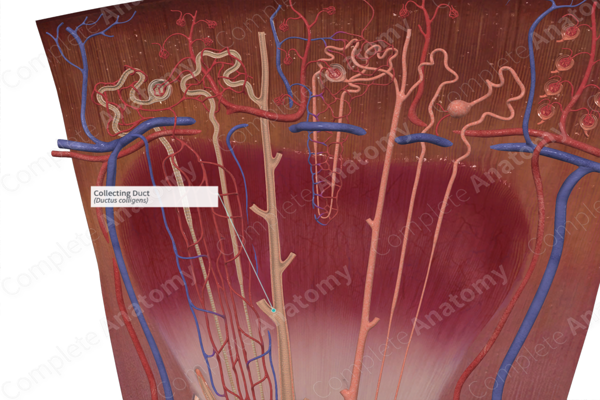
Structure and/or Key Features
The distal convoluted tubules of nephrons drain their renal filtrate into larger vessels known as collecting ducts. Collecting ducts are approximately 20 mm in length and pass from the cortex and medulla to the renal pelvis.
In chronological order, the segments of the collecting duct system include connecting tubules, initial collecting tubules, cortical collecting ducts, medullary collecting ducts, and papillary ducts. The connecting tubules of multiple nephrons merge and drain renal filtrate into cortical collecting ducts.
Cortical collecting ducts are located in the cortex, while medullary collecting ducts travel through the medulla. Cortical collecting ducts have a cuboidal cell lining which gradually becomes taller and forms columnar cells as the ducts descend through the medulla.
The two primary cells of the collecting ducts are principal cells and intercalated cells. Characterized by their pale cytoplasm, principal cells increase in size from the cortex down to the papilla. Intercalated cells have an electron-dense cytoplasm which gives them a darker appearance. These cells are additionally distinguished by the presence of modified microvilli on their luminal surface. Intercalated cells become sparser in the medullary collecting ducts, eventually leaving only principal cells in the papillary collecting ducts (Johnson, Feehally and Floege, 2014).
Anatomical Relations
Collecting ducts extend from the cortex, through the medulla, to the tip of the papilla. The ducts progressively anastomose as they pass from the cortex to the papilla.
Function
The collecting duct plays a pivotal role in fluid electrolyte balance. Specifically, the principal cells of the collecting ducts reabsorb Na+ and water. The darker staining intercalated cells secrete protons and HCO3-. In response to the hormone vasopressin (antidiuretic hormone), the collecting duct reabsorbs water, while in response to the hormone aldosterone, the collecting duct reabsorbs sodium.
List of Clinical Correlates
—Nephrogenic diabetes insipidus
—Liddle’s syndrome
—Pseudohypoaldosteronism type 1
References
Dorland, W. (2011) Dorland's Illustrated Medical Dictionary. 32nd edn. Philadelphia, USA: Elsevier Saunders.
Johnson, R. J., Feehally, J. & Floege, J. (2014) Comprehensive Clinical NephrologyElsevier Saunders.
