Structure/Morphology
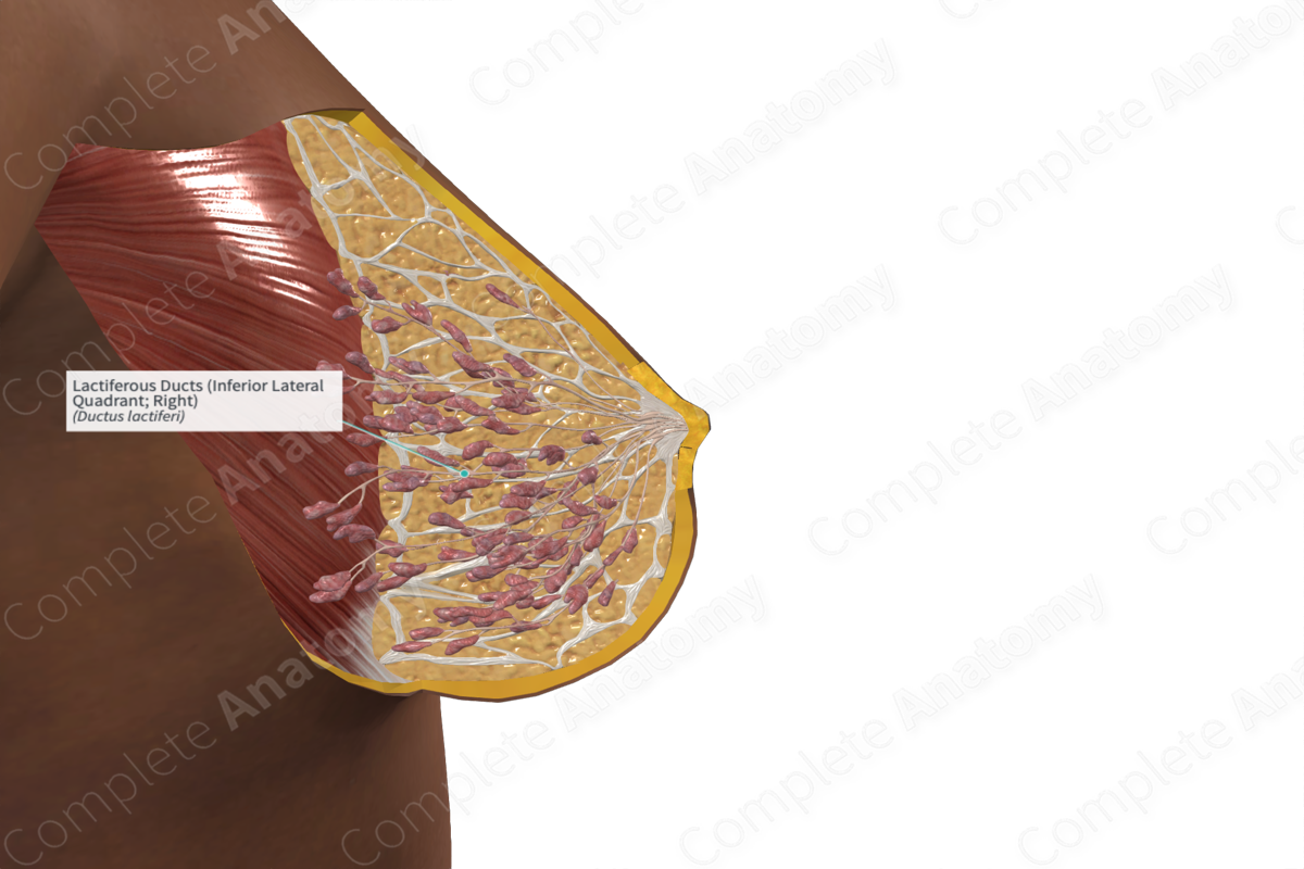
A network of lactiferous ducts connects the lobes of the mammary glands to the nipple for the secretion of milk to the infant.
Milk is synthesized and secreted by the alveoli in the lobules of the mammary glands is secreted into a secondary mammary tubule. The secondary mammary tubules within each lobe unite to form a primary mammary duct, the lactiferous duct. The terminal end of each lactiferous duct has an expansion called the lactiferous sinus, where milk can be stored (Ovalle, Nahirney and Netter, 2013). The sinus narrows again at the base of the nipple and opens onto the surface of the nipple at a small orifice.


Related parts of the anatomy
Function
The lactiferous ducts are responsible for the transport of milk produced in the lobules of the mammary glands to the nipple.
List of Clinical Correlates
- Ductal carcinoma
References
Ovalle, W. K., Nahirney, P. C. and Netter, F. H. (2013) Netter's Essential Histology. ClinicalKey 2012: Elsevier Saunders.
