Quick Facts
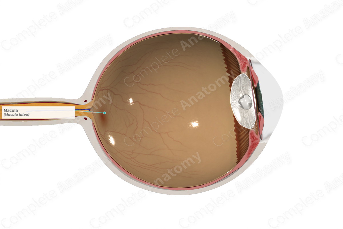
The macula is an irregular yellowish depression on the retina, about 3 degrees wide, lateral to and slightly below the optic disk; it is the site of absorption of short wavelengths of light, and it is thought that its variation in size, shape, and coloring may be related to variant types of color vision (Dorland, 2011).


Related parts of the anatomy
Structure and/or Key Feature(s)
Superior and lateral to the optic disc is a small oval area called the macula or central retina that is characterized on ophthalmoscopic examination by greenish-yellow pigmentation. This region of the retina contains specialized photoreceptors, the cones, responsible for acuity of vision. The fovea centralis is a depression at the center of the macula, which is the site of most acute vision.
Anatomical Relations
The macula is located in the superior temporal retina. It is an oval area of about 5.5 mm in diameter along the long axis. The macula contains the fovea at its center.
Function
The macula is responsible for the central, high-resolution, color vision.
List of Clinical Correlates
—Macular degeneration
References
Dorland, W. (2011) Dorland's Illustrated Medical Dictionary. 32nd edn. Philadelphia, USA: Elsevier Saunders.
