Quick Facts
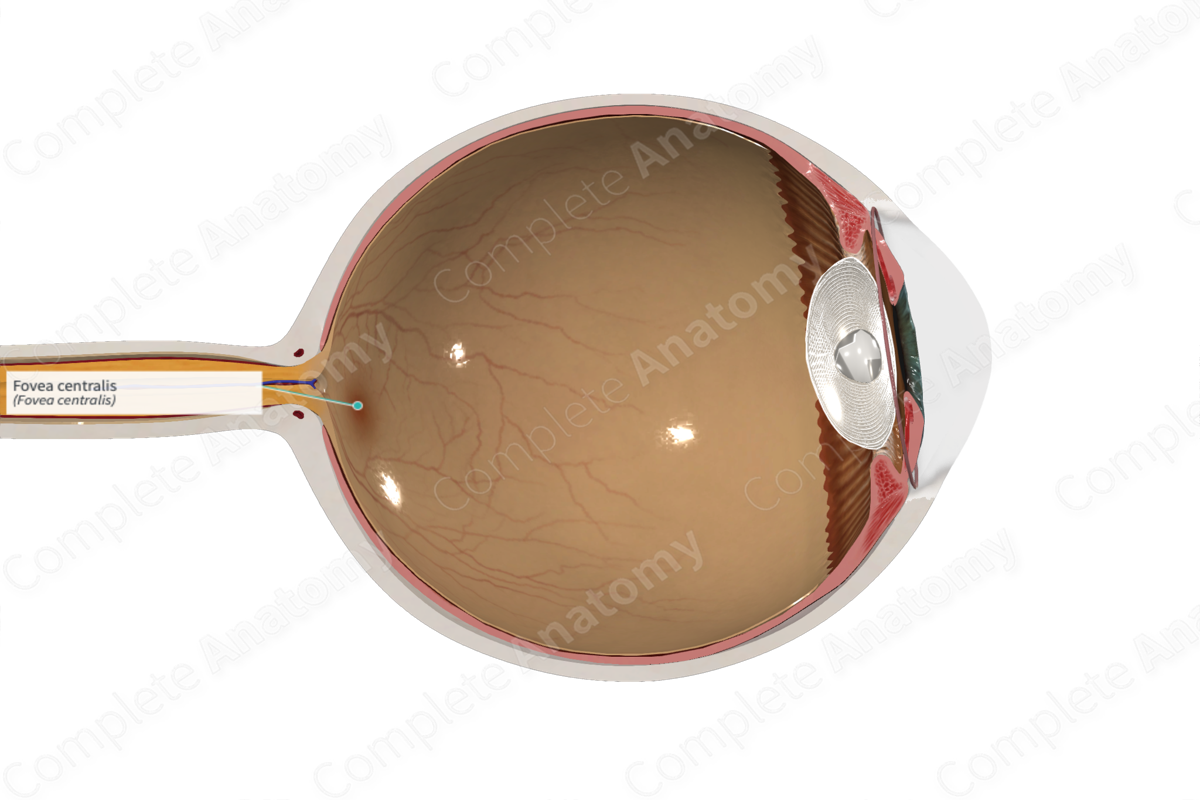
The fovea centralis is a tiny pit, about 1 degree wide, in the center of the macula lutea, which in turn presents an extremely small depression (foveola) containing rodlike elongated cones; it is the area of most acute vision, because here the layers of the retina are spread aside, permitting light to fall directly on the cones (Dorland, 2011).


Related parts of the anatomy
Structure and/or Key Feature(s)
The fovea centralis is a region of the retina that is where the fixation point falls and that produces the highest acuity vision. It is located in the superior temporal retina at the center of the macula. It is a depression surrounded by a mound where the retinal layers internal to the cone photoreceptors are “brushed aside” to be piled up in a surrounding annulus. The fovea centralis contains only cone photoreceptors. These connect to single bipolar cells that connect to single retinal ganglion cells, an arrangement, where there is no convergence of cones provides the maximum resolution of detail, on the order of the diameter of a cone outer segment. The macula has about 4000-5000 cones per millimeter squared, while the fovea centralis has 15,000 cones per millimeter squared (Gupta et al., 2016).
Anatomical Relations
The fovea centralis is found in the superior temporal retina at the center of the macula. Axons from retinal ganglion cells sweep superior and inferior to the fovea on the way to converging on to the optic disc. At the center of the fovea, a region called the foveola, the retina is only as thick as the cone outer segments and cell bodies. This region is avascular, and the metabolic needs of the cells in the fovea are supplied by diffusion from the underlying choroid and sparing the retina from the light perturbing effects of the blood and vasculature.
Function
The fovea is specialized for this highest possible acuity color vision. This is achieved by its many anatomical features including:
—highest density of cells in the retina, purely cone photoreceptors;
—one-to-one connections between photoreceptors, bipolar cells, and retinal ganglion cells;
—and absence of branches of the central retinal artery (in the avascular zone).
References
Dorland, W. (2011) Dorland's Illustrated Medical Dictionary. 32nd edn. Philadelphia, USA: Elsevier Saunders.
Gupta, M. P., Herzlich, A. A., Sauer, T. and Chan, C. C. (2016) 'Retinal Anatomy and Pathology', Dev Ophthalmol, 55, pp. 7-17.
