Structure
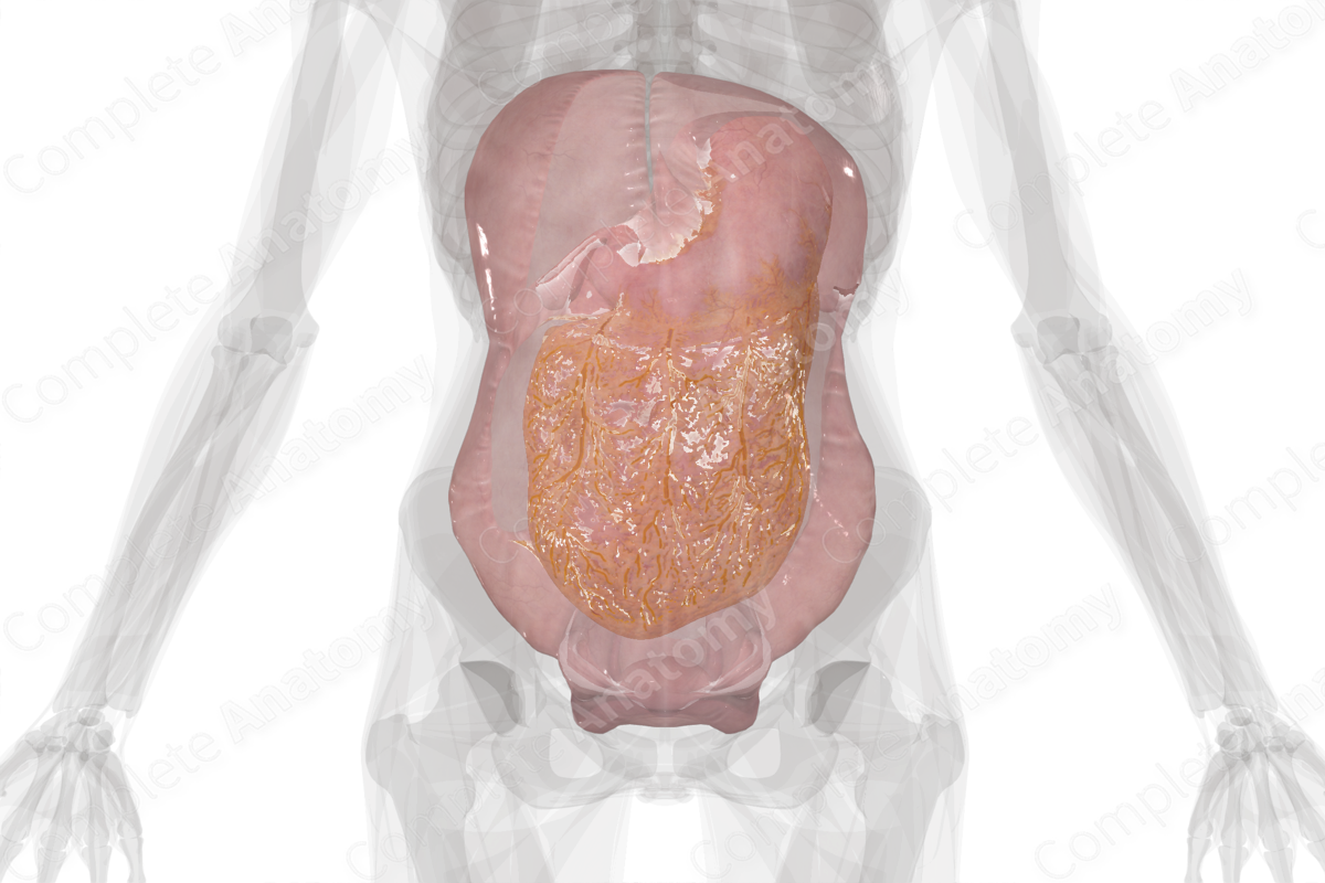
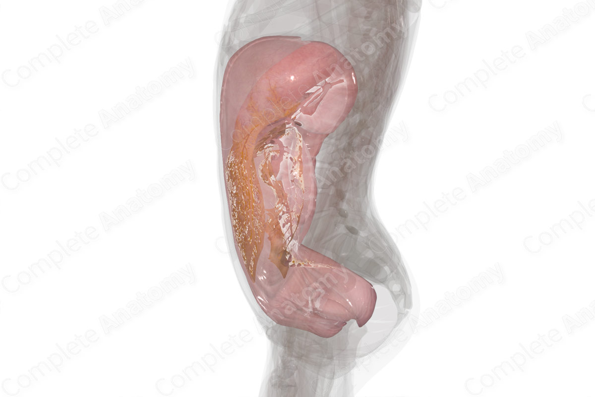
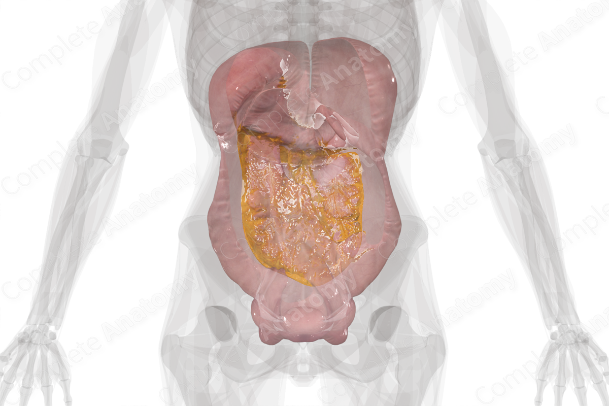
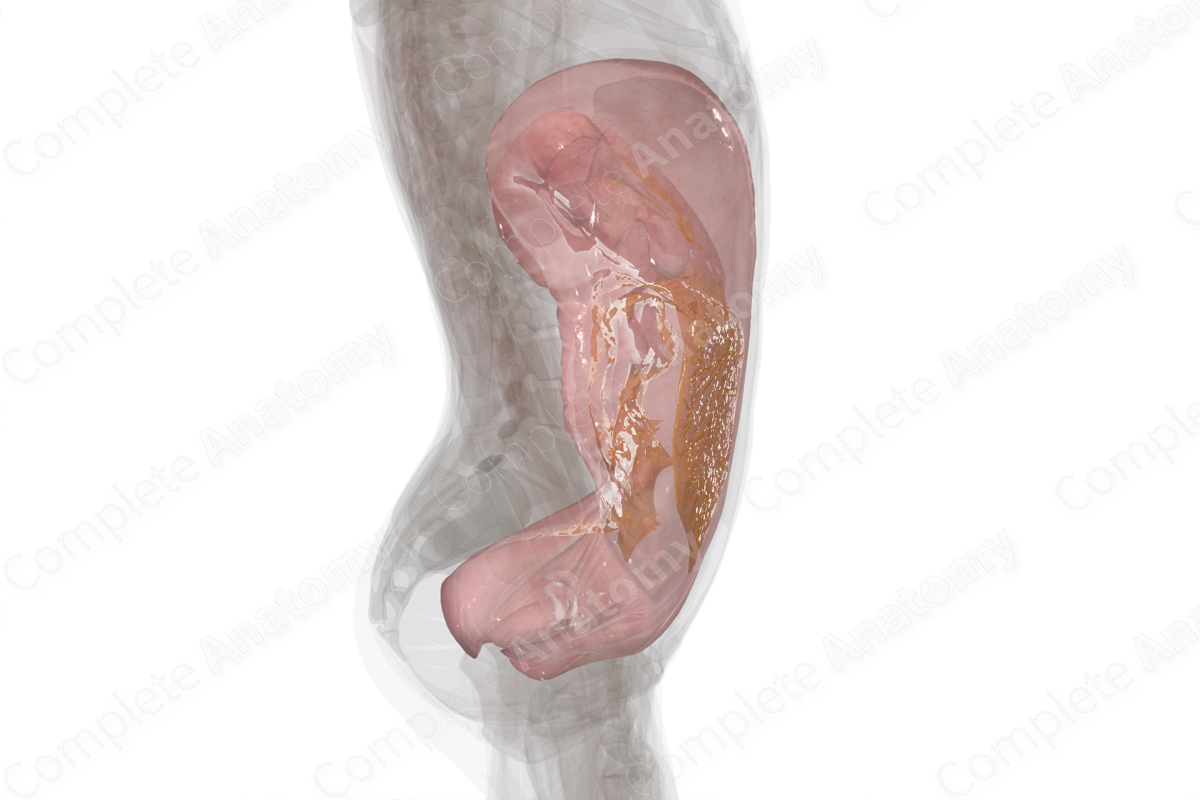
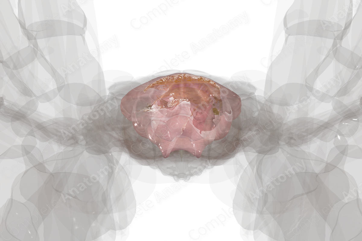
The peritoneum is a complex, continuous serous membrane consisting of a layer of mesothelium and varying degrees of connective and adipose tissue. Visually, it’s largely unremarkable, smooth, and has a lubricated surface due to the presence of peritoneal fluid.
The peritoneum is a single serous membranous closed sac. It consists of two layers, the parietal and visceral peritoneum, that are continuous with each other. The parietal peritoneum lines the internal walls of the abdominal and pelvic cavities. The visceral peritoneum reflects onto the abdominopelvic organs, such as the stomach or uterus.
The majority of abdominal organs principally developed posterior to the peritoneum (retroperitoneally). As these organs enlarged, they pushed into the peritoneal membrane, which enveloped the surface of these organs, creating the visceral layer of peritoneum. These organs continued to move further anteriorly into the peritoneal cavity and after lining the organs, the peritoneal membrane covered the neurovasculature connected to the organs. This double-layer of peritoneum between the parietal and visceral layers of peritoneum is called a mesentery or peritoneal ligament. Mesenteries support organs, limit their movement, and allow for the passage of the neurovasculature between the organs and the abdominopelvic wall.
Organs surrounded by visceral peritoneum are defined as intraperitoneal and are suspended from mesenteries within the peritoneal cavity. Organs that are posterior to the peritoneum are retroperitoneal (e.g., kidneys). Organs that developed in the peritoneum, but eventually settled in place posterior to the peritoneum are secondarily retroperitoneal (e.g., ascending colon).
Function
The peritoneum secretes peritoneal fluid which helps lubricate viscera within the abdominal and pelvic cavities, reducing friction between organs. This is especially true of dynamic organs involved in peristalsis or those that distend due to changes of volume, such as the bladder. Secondly, the peritoneum aids in the immune response as the peritoneal fluid contains various immune cells. The peritoneal fluid actively flows around the peritoneal cavity and is eventually absorbed by lymphatics. The peritoneum, like other tissues, responds to trauma and inflammation, thus helping to protect the viscera (Moore, Dalley and Agur, 2013).
List of Clinical Correlates
—Peritonitis
—Ascites
—Adhesions
References
Moore, K. L., Dalley, A. F. and Agur, A. M. R. (2013) Clinically Oriented Anatomy. Clinically Oriented Anatomy 7th edn.: Wolters Kluwer Health/Lippincott Williams & Wilkins.
Learn more about this topic from other Elsevier products

Peritoneum Anatomy, Peritoneal Cavity, Retroperitoneal Organs

Study peritoneum anatomy and peritoneal cavity with illustrated videos and quizzes. Understand visceral, parietal, retroperitoneal, and subperitoneal organs.
