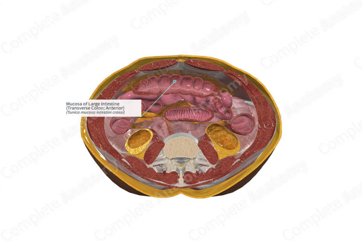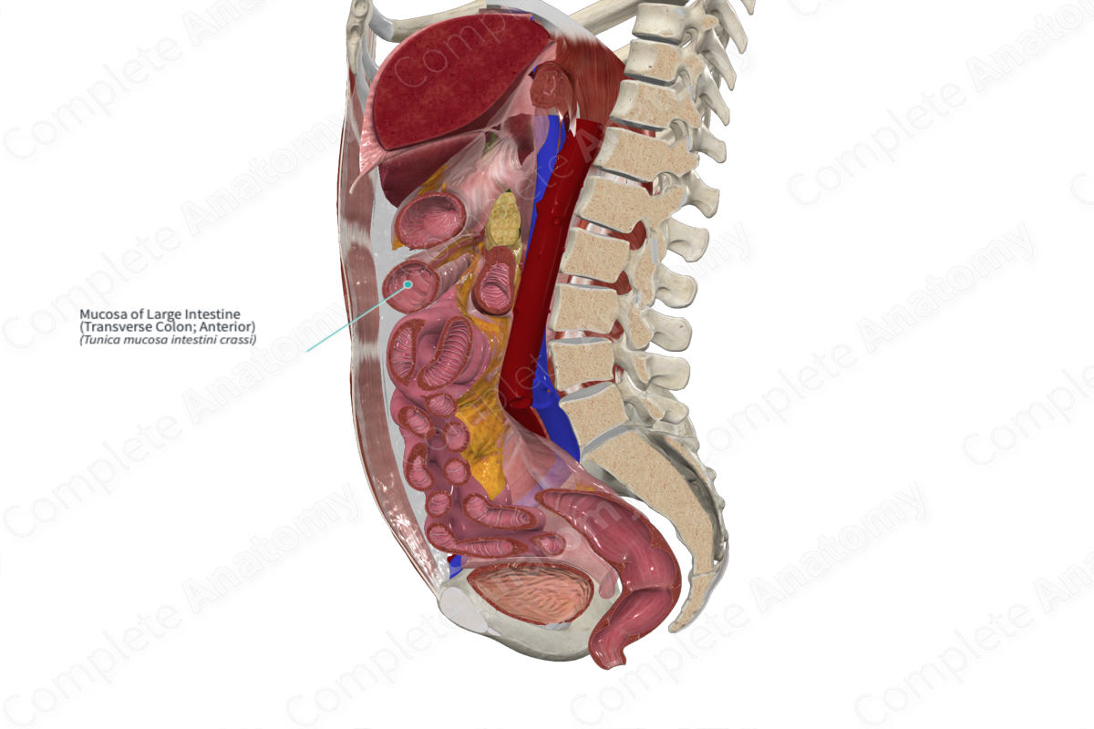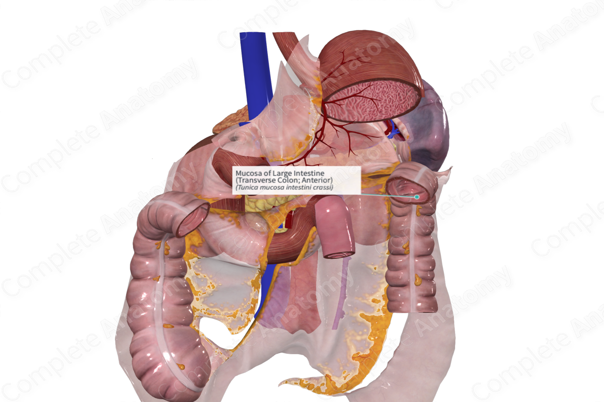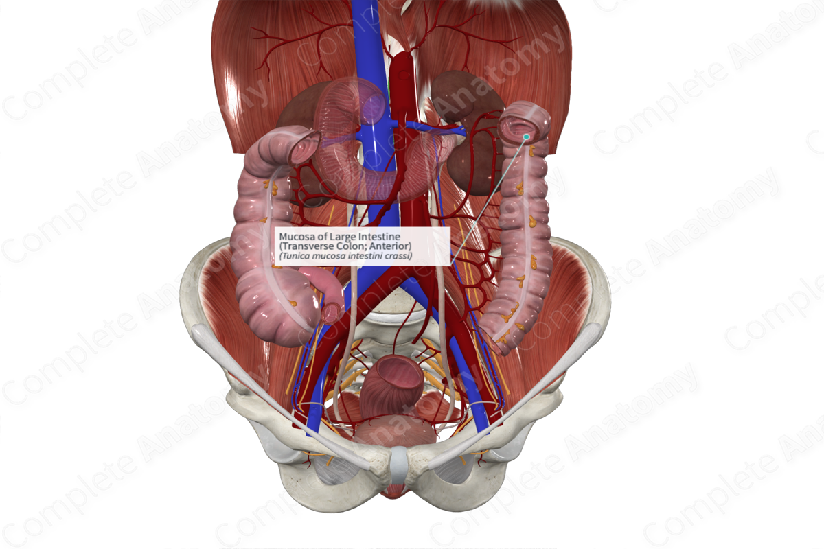
Mucosa of Large Intestine (Transverse Colon; Anterior)
Tunica mucosa intestini crassi
Read moreStructure/Morphology
The mucosa of the large intestine is the inner layer lining the cecum, colon, and rectum. It consists of a simple columnar epithelium that’s in direct contact with the lumen. The epithelium is surrounded by the lamina propria (loose connective tissue) and the muscularis mucosa, a thin layer of smooth muscle that marks the boundary with the submucosa.
Unlike the small intestine, the large intestine mucosa lacks villi. The glands (or crypts of Lieberkühn) contain large amounts of mucin-secreting goblet cells (Standring, 2016).
Related parts of the anatomy
Key Features/Anatomical Relations
The mucosa of the large intestine contains several incomplete infoldings called the semilunar folds, which mark the haustra of the colon. The lower cecum is devoid of these sacculations. They’re identified in the rectum as the transverse rectal folds.
Function
The mucosa of the large intestine is the major site of electrolyte and water reabsorption, as well as the elimination of intestinal contents.
Goblet cells found in the mucosa of the entire large intestine also produce mucus for lubricating the passage of feces.
List of Clinical Correlates
- Ulcerative colitis
- Polyps
- Celiac disease
- Diverticulitis
References
Standring, S. (2016) Gray's Anatomy: The Anatomical Basis of Clinical Practice. Gray's Anatomy Series 41 edn.: Elsevier Limited.
Learn more about this topic from other Elsevier products


