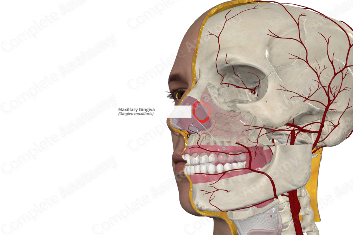
Structure/Morphology
The maxillary gingiva is the region of oral mucosa surrounding the upper dentition and the corresponding alveolar regions of the maxilla. There are two regions of gingivae, the attached and free gingivae. The bulk of the gingival is attached to the alveolar margins of the maxilla. The free gingivae form a thin rim, of approximately 1 mm, around the neck of the teeth (Standring, 2016).
Related parts of the anatomy
Key Features/Anatomical Relations
At the base of the maxilla gingivae, the masticatory mucosa of the gingiva is separated from the lining mucosa by the mucogingival junction. This can be easily differentiated due to the different colors of the different mucosae. The mucosa lining the alveolar surfaces is dark red while the gingival mucosa is pale pink. This is proportional to the level of keratinization and its proximity to the vasculature.
Function
The gingivae act as a barrier against the external environment, such as preventing injury, water loss, and microbial invasion. Additionally, since the gingivae is considered masticatory mucosa, it protects underlying structures from the mechanical stresses of mastication.
List of Clinical Correlates
- Gingivitis
References
Standring, S. (2016) Gray's Anatomy: The Anatomical Basis of Clinical Practice., 41st edition. Elsevier Limited.
Learn more about this topic from other Elsevier products


