Quick Facts
Location: Abdominal cavity.
Arterial Supply: Left colic artery, branch of the inferior mesenteric artery.
Venous Drainage: Left colic vein.
Innervation: Parasympathetic: pelvic splanchnic nerves (S2-S4); Sympathetic: inferior mesenteric plexus; Visceral afferents: L1-L2 spinal ganglia; Enteric innervation.
Lymphatic Drainage: Paracolic nodes to inferior mesenteric lymph nodes.
Related parts of the anatomy
Structure/Morphology
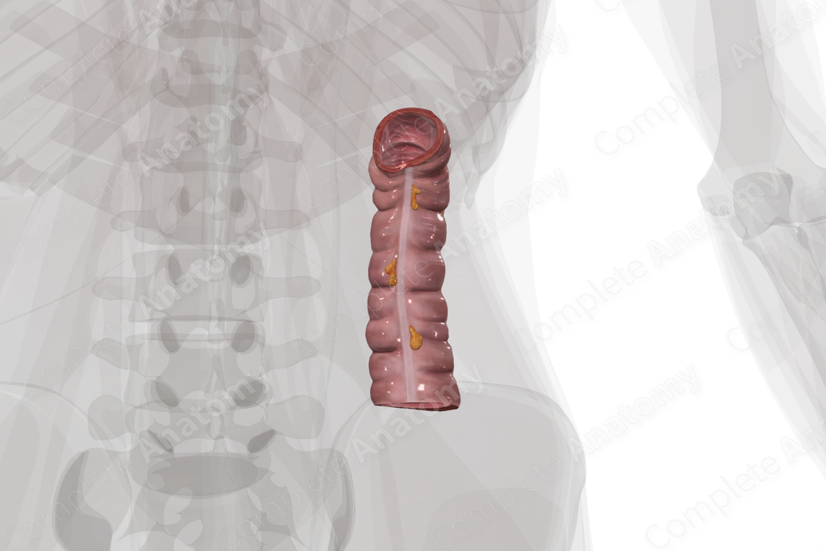
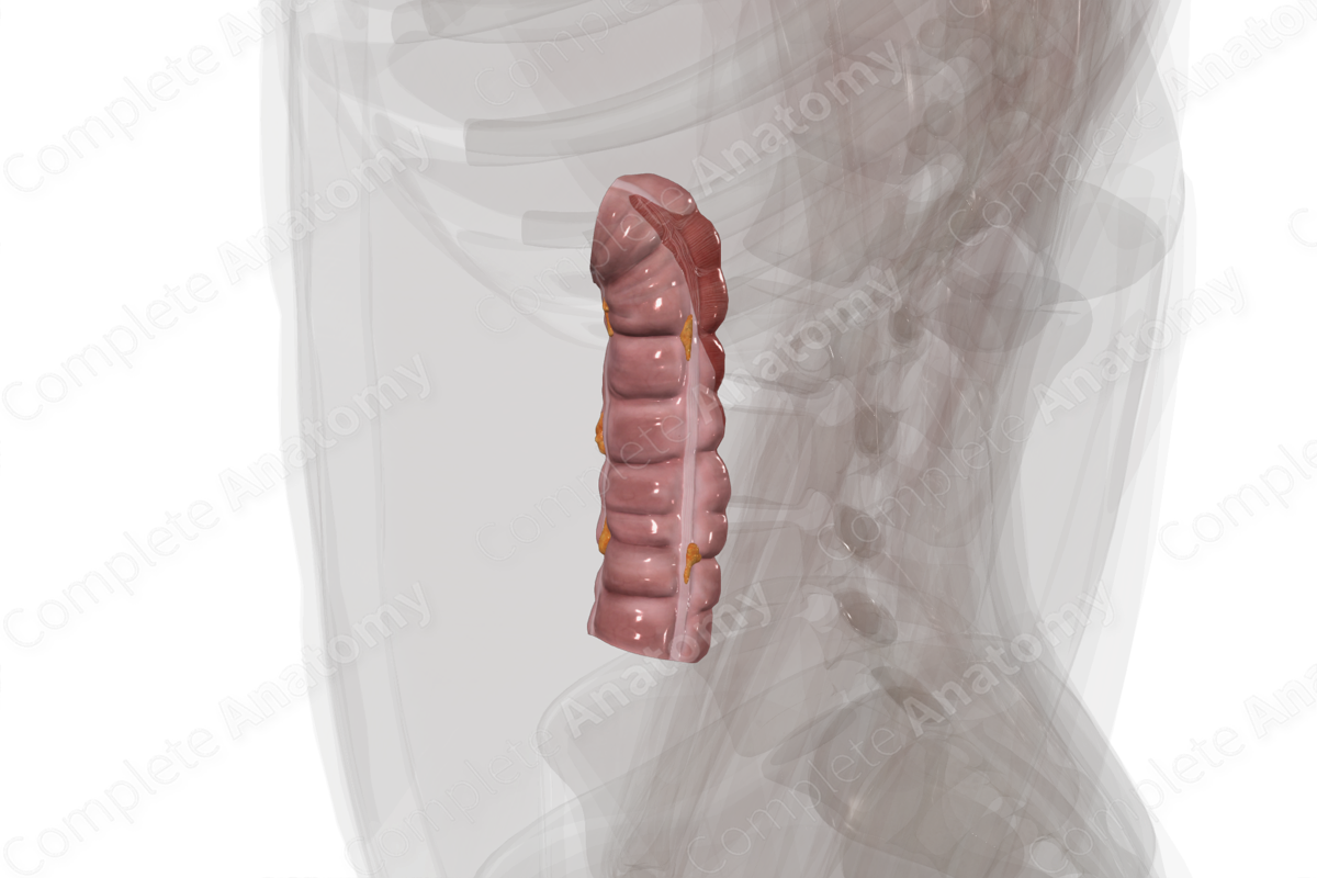
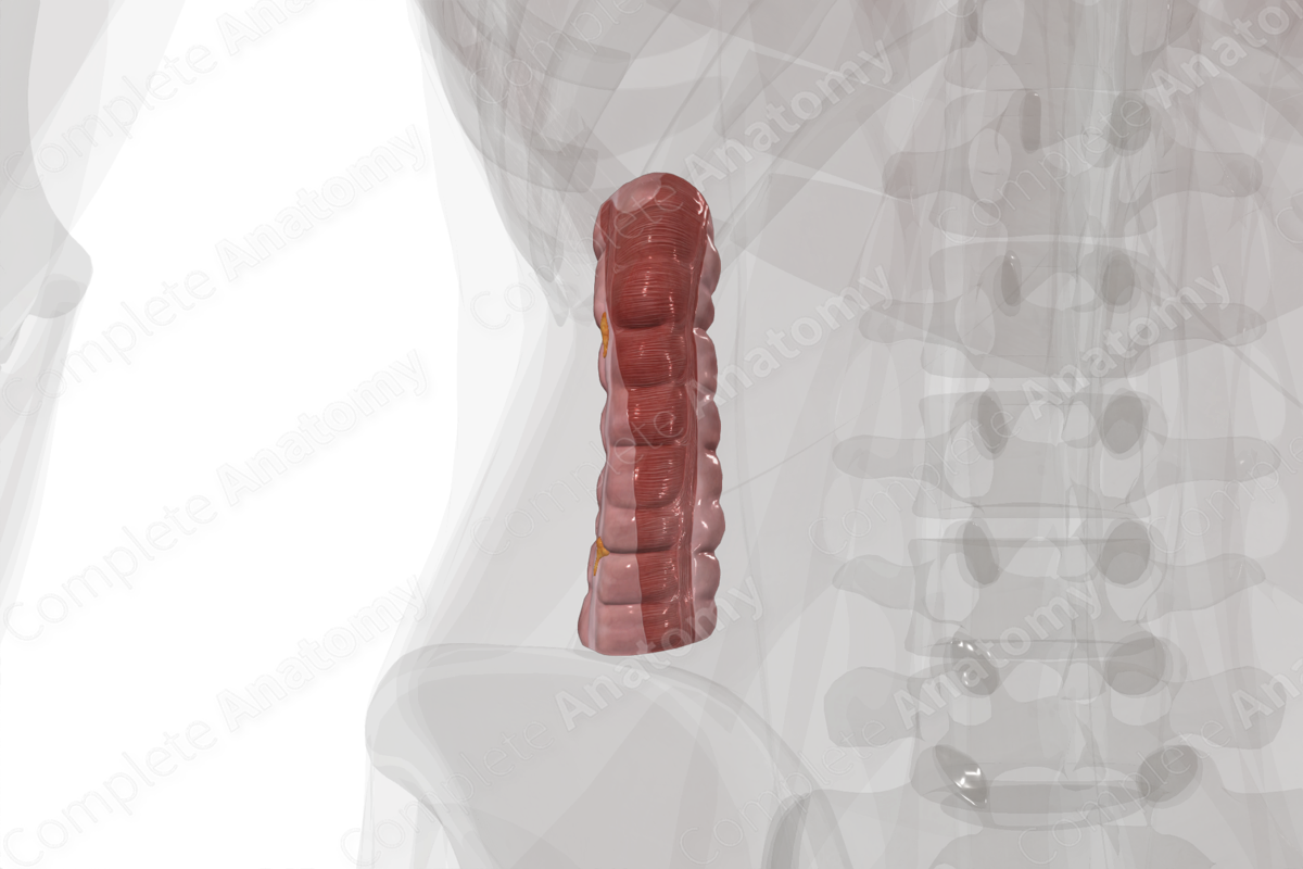
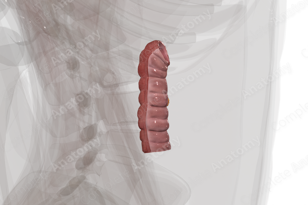
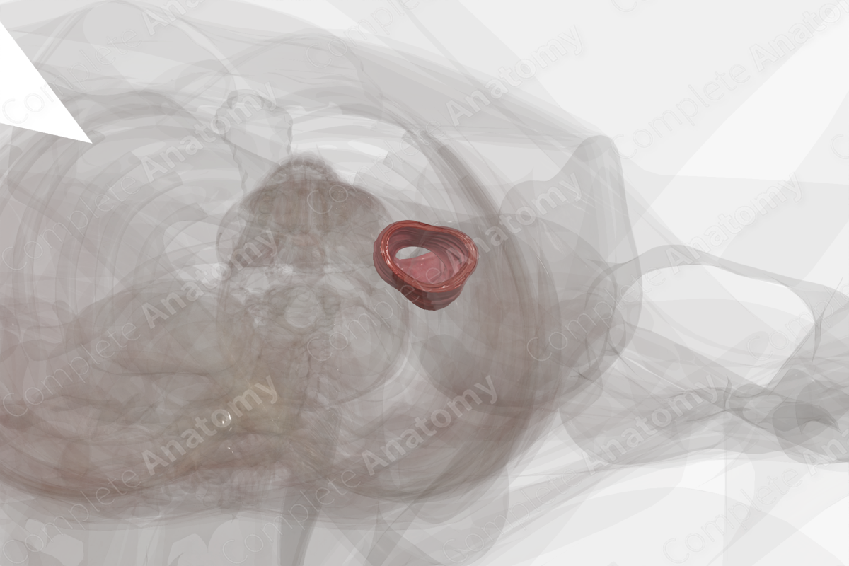
The descending colon is the portion of the large intestine between the splenic flexure and the sigmoid colon.
The cross-sectional microarchitecture of the descending colon demonstrates mucosa, submucosa, and muscular (inner circular and outer longitudinal) layers with a thin outer serosal covering (Standring, 2016).
As with the rest of the large intestine, the longitudinal muscle arrangement of the descending colon contains three muscular bands called teniae coli. Contraction of the teniae coli shortens the descending colon to create sacculations or bulges which are called haustra. Between the haustra are the semilunar folds. These promote churning of chyme and increase the surface area of mucosa for absorption (Standring, 2016).
Additionally, small pockets of fat called the omental appendices project from the external (non-mesenteric) surface of most of the large intestine.
The descending colon is part of the hindgut, which helps explain the patterns of vascularization, innervation, and lymphatic drainage of this tissue.
Anatomical Relations
The proximal descending colon begins at the splenic flexure where the colon bends inferiorly and is anchored to the diaphragm by the phrenicocolic ligament. It runs retroperitoneally, inferiorly down the posterior abdominal wall passing anterior to the left kidney. The small intestine lies medial to the descending colon, while the left lateral paracolic gutter sits laterally. Upon reaching the left lower pelvis, the descending colon bends to the right and becomes the sigmoid colon.
Function
The descending colon absorbs most of the remaining water and essential vitamins still present in the waste material. Most other nutrients will have previously been absorbed.
The descending colon also functions as a storage space for the solid fecal material. It contracts peristaltically to propel feces into the sigmoid colon and rectum during defecation.
Arterial Supply
The descending colon is primarily supplied by the left colic artery, a branch of the inferior mesenteric artery.
A significant anastomotic arterial “circle” known as the marginal artery of the colon (of Drummond) allows blood from an artery serving one area of the colon to pass to territories normally served by a different artery. This potentially allows blood from the middle colic artery to serve the descending colon.
Venous Drainage
The venous drainage is into the portal circulation. The pattern largely parallels the arterial supply, with blood from the left colic vein draining to the inferior mesenteric vein, the splenic vein, the portal vein, and into the liver.
Innervation
Innervation of the descending colon includes the enteric nervous system (sensory and motor), the autonomic nervous system (sympathetic and parasympathetic), and extrinsic sensory innervation (visceral afferents) (Standring, 2016). The descending colon is part of the hindgut, and the patterns of innervation reflect this.
Parasympathetic innervation to the descending colon is supplied by pelvic splanchnic nerves from S2-S4.
Sympathetic innervation is derived from the sympathetic chain or the aortic plexus. The postsynaptic neurons are found in the inferior mesenteric ganglia and reach the descending colon via the inferior mesenteric plexus.
Visceral afferent nerves from the descending colon travel back to the CNS along the lumbar and pelvic splanchnic nerves.
The enteric system consists of two plexuses of densely packed small neurons. Meissner’s plexus lies in the submucosal layer and Auerbach’s myenteric plexus lie between the outer longitudinal and inner circular smooth muscle layers. These systems of nerves control mucosal and peristaltic function.
Lymphatic Drainage
Lymphatic drainage from the descending colon parallels the arterial supply. Paracolic nodes drain into intermediate colic nodes, then to inferior mesenteric lymph nodes. Ultimately, all lymph is drained via the cisterna chyli and the thoracic lymphatic duct.
List of Clinical Correlates
- Colitis
- Colonoscopy
- Diverticulitis
- Colorectal Cancer
References
Standring, S. (2016) Gray's Anatomy: The Anatomical Basis of Clinical Practice. Gray's Anatomy Series 41 edn.: Elsevier Limited.
Learn more about this topic from other Elsevier products

