Quick Facts
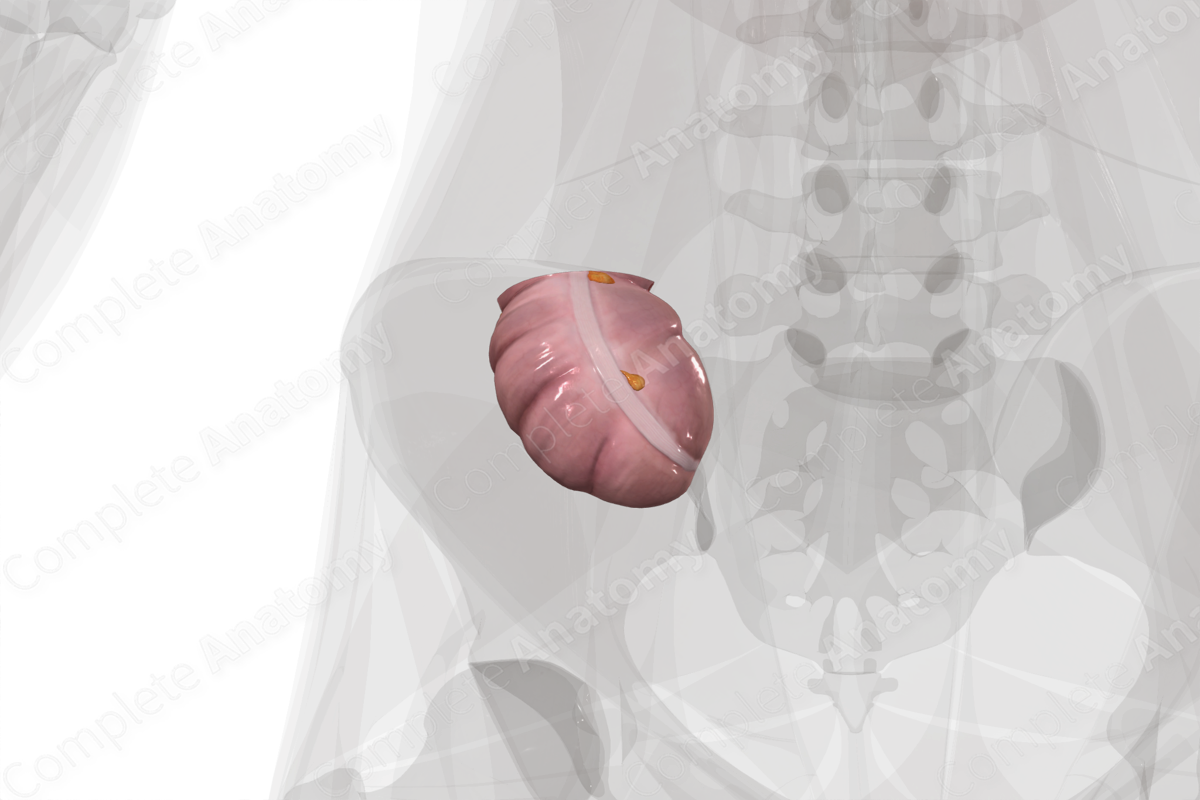
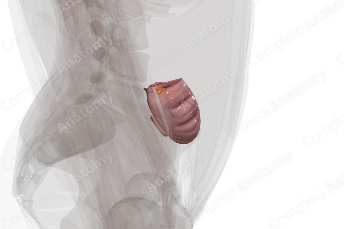
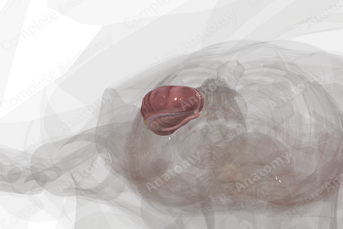
Location: Pelvic cavity.
Arterial Supply: Ileocolic artery, branch of the superior mesenteric artery.
Venous Drainage: Ileocolic vein to the superior mesenteric vein and portal system.
Innervation: Parasympathetic: vagus nerve (CN X); Sympathetic: celiac and superior mesenteric ganglia; Visceral afferents: spinal ganglia of T5-T9; Enteric innervation.
Lymphatic Drainage: Ileocolic nodes to central superior mesenteric nodes.

Related parts of the anatomy
Structure/Morphology
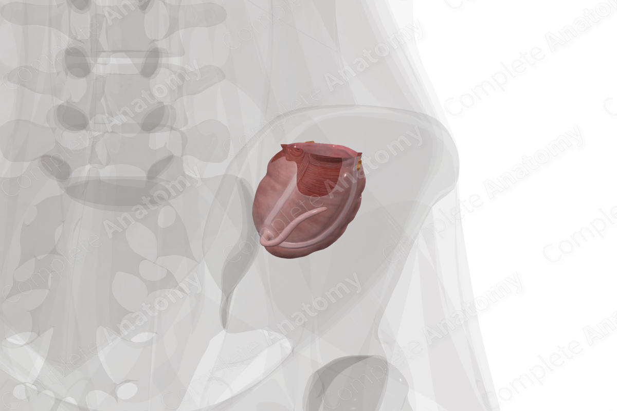
The cecum forms the first part of the large intestine.
The cross-sectional microarchitecture of the cecum demonstrates mucosa, submucosa, and muscular (inner circular and outer longitudinal) layers with a thin outer serosal covering.
The longitudinal muscle arrangement of both the cecum and colon is distinct. It contains three muscular bands called teniae coli. When the teniae coli contract, the cecum and colon are shortened leading to localized regions of expansion and constriction. These sacculations of the large intestinal wall are called haustra. Additionally, small pockets of fat called the omental appendices project from the external (non-mesenteric) surface of most of the large intestine.
The cecum is part of the midgut. This developmental distinction is important in understanding its patterns of vascularization, innervation and lymphatic drainage.
Anatomical Relations
The cecum is the first part of the large intestine and is continuous with the small intestine at the ileocecal junction.
The cecum lies in the right iliac fossa of the lesser pelvis and extends inferiorly from the ileocecal junction towards the inguinal ligament. It is intraperitoneal, and the appendix is a diverticular outgrowth of the inferior cecum.
Slightly superior to the ileocolic junction, the cecum transitions to the ascending colon at a constriction called the cecocolic junction.
Function
The cecum receives soft, water-rich wastes from the ileum and can store a considerable volume of chyme due to its distensible nature. It's also the site of attachment for the appendix, a vestigial structure containing lymphoid tissue.
The cecum absorbs water, salts, vitamins, and minerals from the waste material, and secretes mucus from its epithelium.
Arterial Supply
The cecum is supplied by anterior and posterior cecal arteries, which are branches of the ileocolic artery, and serve the anterior and posterior surfaces, respectively. The ileocolic artery is in turn a branch of the superior mesenteric artery.
Venous Drainage
The venous drainage is into the portal circulation. The pattern largely parallels the arterial supply with blood draining via the ileocolic vein to the superior mesenteric vein, the portal vein, and ultimately the liver.
Innervation
Innervation of the cecum includes the enteric nervous system (sensory and motor), autonomic nervous system (sympathetic and parasympathetic), and extrinsic sensory innervation (visceral afferents) (Standring, 2016).
Sympathetic innervation is derived from the sympathetic chain or the aortic plexus. The cecum is part of the midgut and is supplied by postganglionic neurons from the celiac and superior mesenteric ganglia.
The vagus nerve (CN X) provides parasympathetic innervation to the cecum.
Visceral afferent nerves from the midgut ascend in tandem with the vagus nerve and greater splanchnic nerve.
The enteric system consists of two plexuses of densely packed small neurons. Meissner’s plexus lies in the submucosal layer and Auerbach’s myenteric plexus lie between the outer longitudinal and inner circular smooth muscle layers. These systems of nerves control mucosal and peristaltic function.
Lymphatic Drainage
Lymphatic drainage from the cecum parallels the arterial supply and drains into the superior mesenteric nodes. Generally, lymph flows from cecal and ileocolic nodes to the central superior mesenteric nodes. Ultimately lymph drains into the cisterna chyli (Standring, 2016).
List of Clinical Correlates
- Colitis
- Diverticulitis
- Cecal volvulus
References
Standring, S. (2016) Gray's Anatomy: The Anatomical Basis of Clinical Practice. Gray's Anatomy Series 41 edn.: Elsevier Limited.
Learn more about this topic from other Elsevier products

