Anatomical Relations
The palmar aponeurosis radiates into the fingers and covers the soft tissue and long flexor tendons of the hand. The median nerve and the superficial palmar arch travels deep to the aponeurosis to reach their targets, while the palmar cutaneous branch of the ulnar nerve travels above the palmar aponeurosis. The flexor tendons pass through short channels formed between the palmar aponeurosis and the heads of the metacarpal bones. The palmar digital arteries and nerves pass between the four digital slips of the aponeurosis to reach the fingers.


Related parts of the anatomy
Structure
The palmar aponeurosis is a strong, triangular membrane covering the tendons and muscles on the palmar surface of the hand.
The apex of the longitudinal fibers is continuous with palmaris longus, when present, or is anchored to the flexor retinaculum of the wrist. The fibers run distally producing four longitudinal bundles, which travel to the index, middle, ring, and little fingers. A less well-defined bundle passes to the thumb. Distal to the transverse fibers of the palmar aponeurosis, the longitudinal fibers divide into three layers.
—The first, superficial longitudinal fibers are inserted superficially into the skin of the distal palm and the base of the fingers.
—The second, middle longitudinal fibers pass deep to the superficial transverse metacarpal ligament and into the fingers where they are continuous with the lateral digital sheaths.
—The third, deepest layer of longitudinal fibers penetrate the deep traverse metacarpal ligament to pass around the sides of the metacarpophalangeal joint and attach to the metacarpal bone, proximal phalanx, and extensor tendon.
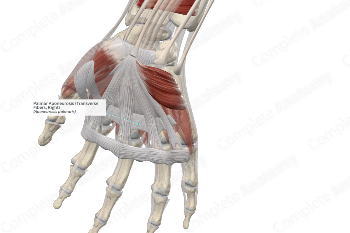
The transverse fibers of the palmar aponeurosis are the deepest layer of the palmar fascia. They lie proximal to the distal palmar crease in a band approximately 2 cm wide. The anterior fibers of the flexor tendon blend with the anterior fibers of the flexor tendon sheaths. Vertical fibers connect the palmar aponeurosis to the thenar and hypothenar eminences.
Function
The palmar aponeurosis acts as the insertion point for the tendon of palmaris longus and is the origin point for palmaris brevis. The transverse fibers support the webs between the fingers. As it attaches to the skin, it improves the grip of the hand and helps protect the under-lying tendons.
List of Clinical Correlates
—Dupuytren contracture
References
Standring, S. (2016) Gray's Anatomy: The Anatomical Basis of Clinical Practice. Gray's Anatomy Series 41st edn.: Elsevier Limited.
Learn more about this topic from other Elsevier products

