Structure
Bursae are sac-like structures, with an inner synovial membrane, that produces a thin film of synovial fluid. They aid in reducing friction between moving tissues of the body, such as between tendon and bone, ligament and bone, tendons and ligaments, and between muscles.
Inflammation of the bursa is known as bursitis. If the inflammation is due to injury or strain, it is known as aseptic bursitis. However, if the inflammation is caused by infection, it is known as septic bursitis.
Related parts of the anatomy
Anatomical Relations
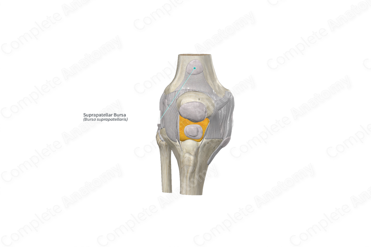
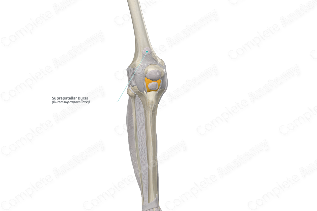
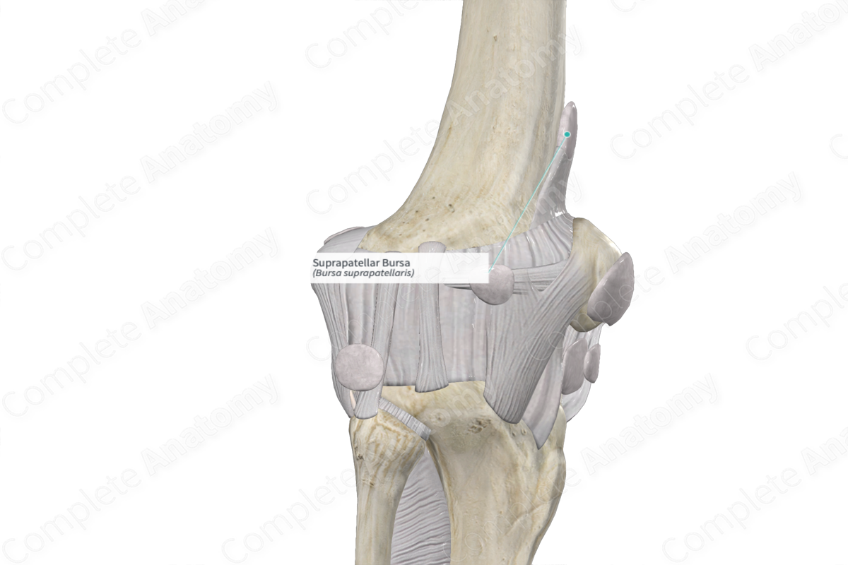
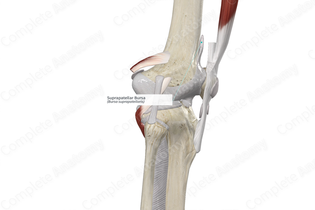
The suprapatellar bursa is located above the patella, between the tendon of the quadriceps femoris muscle and the distal end of the femur. It usually communicates with the knee joint cavity, as the suprapatellar septum separating the suprapatellar bursa from the knee joint cavity involutes at three months of age, leading to the formation of a single joint space. In some cases, the suprapatellar synovial plica, a remnant of the suprapatellar septum, can lead to complete or incomplete isolation of the bursa from the knee joint cavity (Yamamoto et al., 2003).
The articularis genu muscle is a small muscle which lies underneath the vastus intermedius muscle. It inserts into the suprapatellar bursa superiorly and prevents the bursa from collapsing into the joint cavity.
Function
The suprapatellar bursa aids in the movement of the quadriceps tendon over the distal end of the femur.
References
Yamamoto, T., Akisue, T., Marui, T., Hitora, T., Nagira, K., Mihune, Y., Matsui, N., Yoshiya, S. and Kurosaka, M. (2003) 'Isolated suprapatellar bursitis: Computed tomographic and arthroscopic findings', Arthroscopy, 19(2), pp. E10.
Learn more about this topic from other Elsevier products


