Subcutaneous Infrapatellar Bursa (Right)
Bursa subcutanea infrapatellaris
Read moreStructure
Bursae are sac-like structures, with an inner synovial membrane, that produces a thin film of synovial fluid. They aid in reducing friction between moving tissues of the body, such as between tendon and bone, ligament and bone, tendons and ligaments, and between muscles.
Inflammation of the bursa is known as bursitis. If the inflammation is due to injury or strain, it is known as aseptic bursitis. However, if the inflammation is caused by infection, it is known as septic bursitis.


Related parts of the anatomy
Anatomical Relations
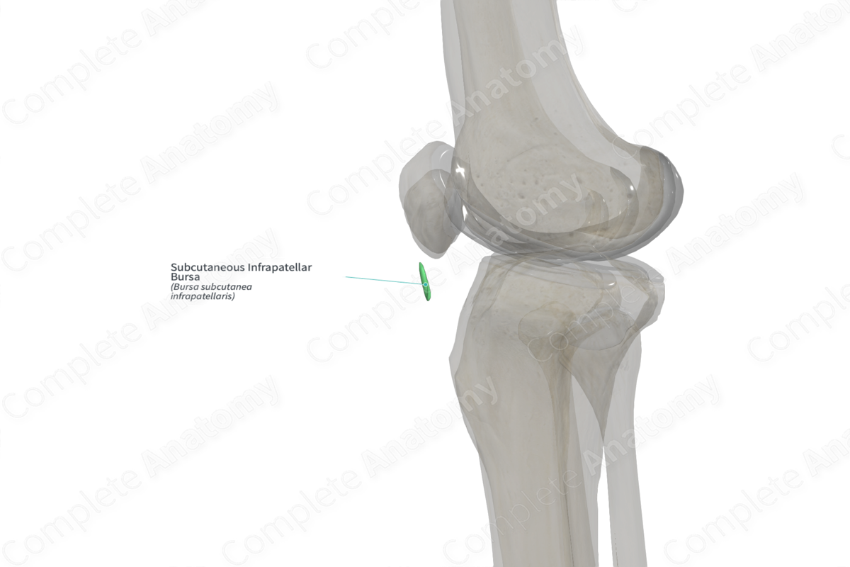
The subcutaneous infrapatellar bursa is a closed sac of synovial fluid found anterior to the patellar ligament, a ligament sitting between the skin and tibial tuberosity. It measures approximately 19.5 mm craniocaudally and 21.2 mm in the mediolateral plane. While the mean size in the anteroposterior plane is approximately 2.2 mm, the transverse limits of the bursa do not extend beyond the patellar tendon. However, in a few cases, the lateral connections between the superficial and deep infrapatellar bursae have been documented along the lateral margins of the patellar tendon (Viegas et al., 2007).
Function
The subcutaneous infrapatellar bursa reduces friction between the patellar ligament and the overlying skin.
List of Clinical Correlates
—Clergyman’s knee
References
Viegas, F. C., Aguiar, R. O., Gasparetto, E., Marchiori, E., Trudell, D. J., Haghighi, P. and Resnick, D. (2007) 'Deep and superficial infrapatellar bursae: cadaveric investigation of regional anatomy using magnetic resonance after ultrasound-guided bursography', Skeletal Radiol, 36(1), pp. 41-6.
Learn more about this topic from other Elsevier products

