
Unfortunately we don't fully support your browser. If you have the option to, please upgrade to a newer version or use Mozilla Firefox, Microsoft Edge, Google Chrome, or Safari 14 or newer. If you are unable to, and need support, please send us your feedback.

Elsevier
Publish with us
Right Superior Phrenic Vein
Cardiovascular System

Right Superior Phrenic Vein

Vena phrenica superior dextra

Read more

Quick Facts

Origin: Union of small tributaries.

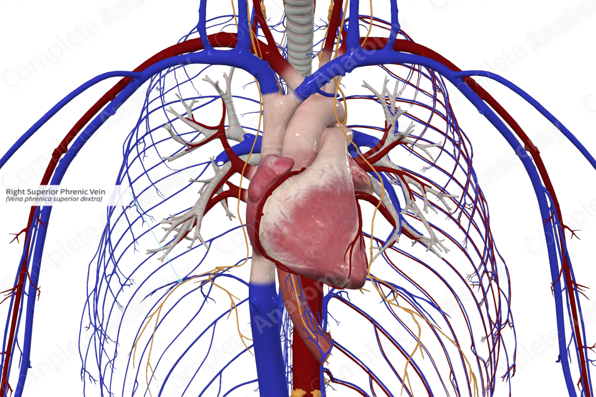
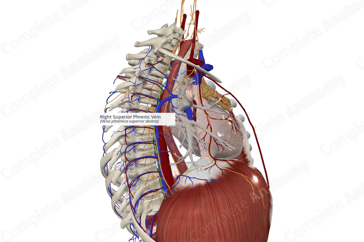
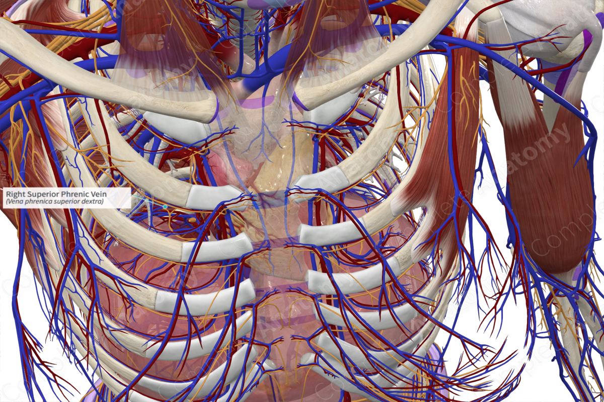
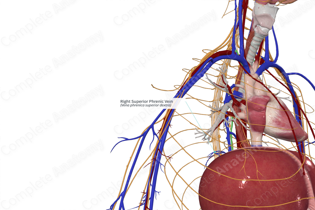
Course: Along superior surface of respiratory diaphragm.

Tributaries: None.

Drainage: Superior surface of diaphragm.

Complete Anatomy
The world's most advanced 3D anatomy platform
Try it for Free

Origin

The superior phrenic veins are formed by the union of small venules on the superior surface of the respiratory diaphragm.

Course

The superior phrenic veins course along the superior surface of the respiratory diaphragm and are accompanied by the superior phrenic arteries. They descend posteriorly to unite with the hemiazygos and azygos veins.

Tributaries

There are no named tributaries; however, they do form an anastomosis with the musculophrenic and pericardiacophrenic veins to ensure constant drainage of the diaphragm.

Structures Drained

The superior phrenic veins drain the vertebral portion of the respiratory diaphragm.

Learn more about this topic from other Elsevier products

Vein

ScienceDirect image

Vein is a secreted ligand for the Drosophila EGFR receptor homolog (EGFR), and EGFR-mediated activation of the Ras/MAP kinase pathway is essential for longitduinal glial cell survival (Hidalgo et al., 2001).

Explore on ScienceDirect(opens in new tab/window)

Complete Anatomy

The world's most advanced 3D anatomy platform

Complete Anatomy