Quick Facts
Origin: Union of pulmonary segmental veins.
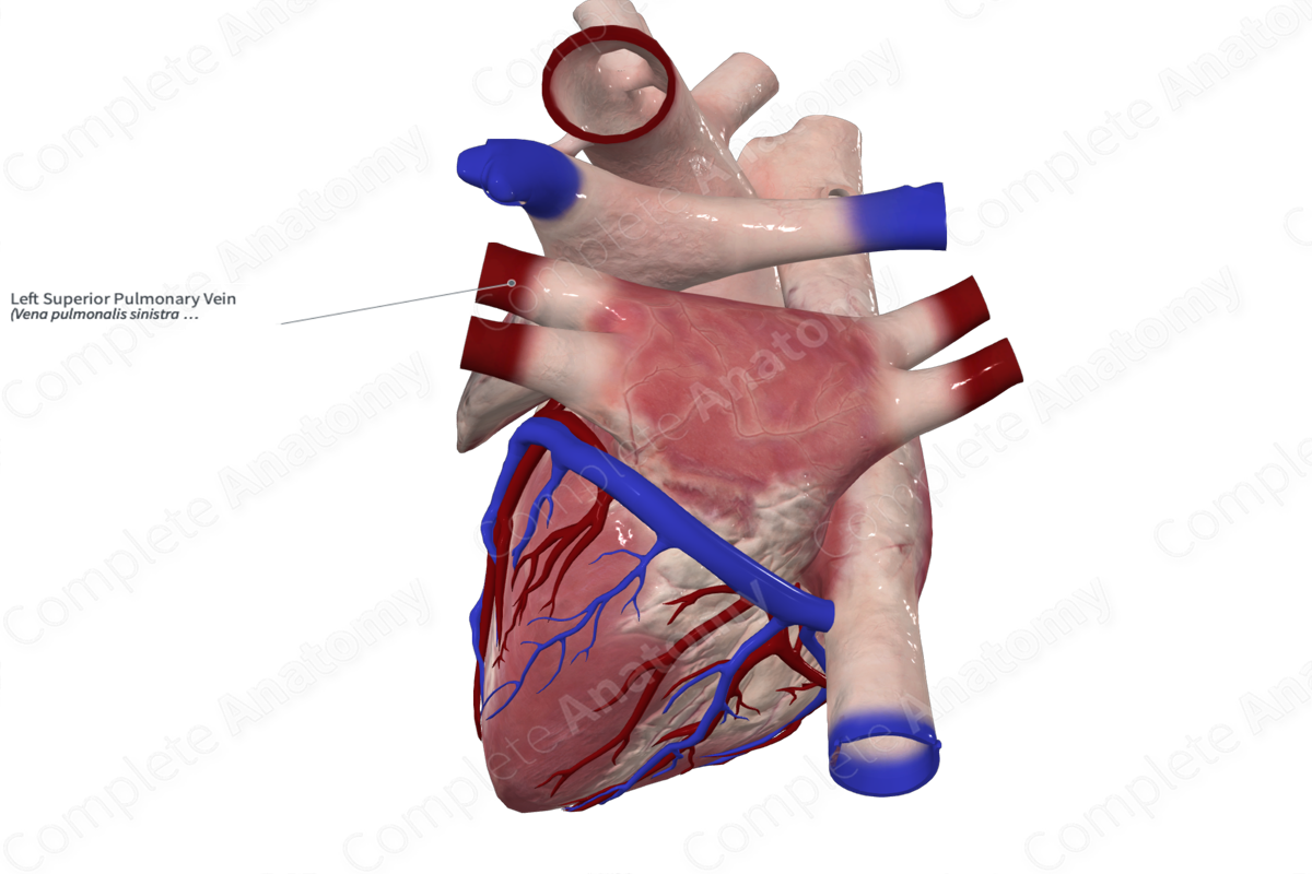
Course: From the left lung to the left posterosuperior aspect of the left atrium.
Tributaries: Anterior, apicoposterior, and lingular veins.
Drainage: Superior lobe of left lung.


Origin
The left superior pulmonary vein is formed in the parenchyma of the superior lobe of the left lung.
Course
As the left superior pulmonary vein exits the hilum of the left lung, it sits anterior to left main bronchus and anteroinferior to the left pulmonary artery. As it travels medially, the left superior pulmonary vein crosses anterior to the descending thoracic aorta, pierces the fibrous pericardium, and drains into the posterosuperior aspect of the left atrium.
Tributaries
The anterior, apicoposterior, and lingular veins unite to form the left superior pulmonary vein.
Structures Drained
The left superior pulmonary vein is responsible for the venous drainage of oxygenated blood from the superior lobe of the left lung.
List of Clinical Correlates
- Pulmonary vein atresia
- Anomalous pulmonary venous connection
Learn more about this topic from other Elsevier products

