Quick Facts
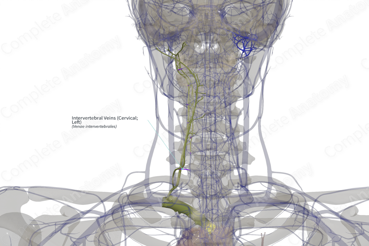
Origin: Union of the anterior and posterior internal venous plexuses and radicular veins.
Course: Pass through the intervertebral foramen.
Tributaries: Anterior and posterior segmental medullary veins and the vertebral venous plexus.
Drainage: Spinal cord, subarachnoid space, epidural space, and vertebral bodies.
Related parts of the anatomy
Origin
The intervertebral veins are formed by the union of the radicular veins and the internal vertebral venous plexuses, meaning it collects blood draining from most of the vertebral column and spinal cord.
Course
The intervertebral veins pass laterally through the intervertebral foramina and accompany the spinal nerves to reach the vertebral vein in the neck, the posterior intercostal veins in the thorax, and the lumbar and lateral sacral veins caudally.
Tributaries
The radicular veins drain into the intervertebral veins, along with the internal and external vertebral venous plexuses. While the latter have collateral flow through the deep cervical, azygos, lumbar, and lateral sacral veins, there is commonly substantial flow through the intervertebral veins.
Structures Drained
The intervertebral veins drain the spinal cord, subarachnoid and epidural spaces, and the vertebrae.
List of Clinical Correlates
- Posterolateral disk herniation
Learn more about this topic from other Elsevier products


