Morphology/Structures
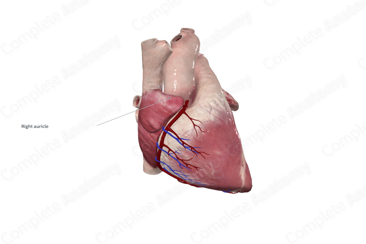
The right auricle is a broad, triangular muscular pouch that is visible on the exterior of the heart and overlaps the ascending aorta. Its internal surface, like the anterior walls of the atria, contains pectinate muscles which contribute to its roughened appearance.


Key Features/Anatomical Relations
The outer anterior wall of the right auricle sits posterior to the right upper sternum. Its outer anterolateral wall lies within the cardiac impression of the right lung. The hilum of the right lung sits both posteriorly and superiorly to the right auricle.
Function
The right auricle is a remnant of the fetal right atrium. The auricles can relieve high atrial pressure by increasing the atrial capacity at times of stress, acting as overflow vessels.
Learn more about this topic from other Elsevier products

