
Morphology/ Structure:
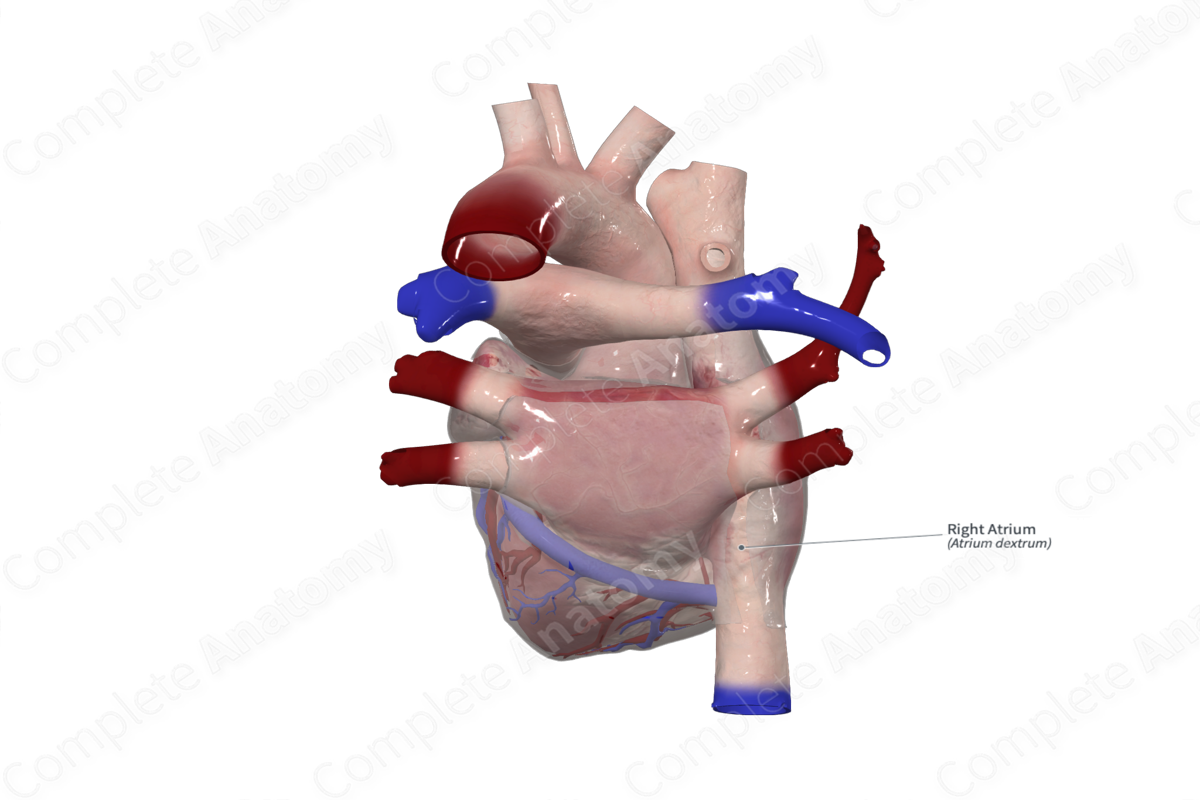
The right atrium sits anterior, inferior and to the right of the left atrium. It forms a portion of the upper anterior (sternocostal) surface and a portion of the right pulmonary surface of the heart.
The right atrium has thin muscular walls as it functions under low pressure. The posterior wall is smooth while the anterior wall of the right atrium is ridged due to the presence of pectinate muscles. These are roughly parallel bars of muscle that run in an anterolateral direction from the crista terminalis.
The right auricle sits over the base of the ascending aorta and contains pectinate muscle, and therefore, has a ridged internal appearance.
The right atrium receives deoxygenated blood via the superior vena cava, inferior vena cava, and the coronary sinus. The superior vena cava has no valve; however, the inferior vena cava is covered by the Eustachian valve, which runs into the Eustachian ridge. The coronary sinus is covered by the Thebesian valve. The outflow tract consists of the smooth-walled vestibule and marks the location of the right atrioventricular valve (or tricuspid valve).


Related parts of the anatomy
Key Features/ Anatomical Relations:
The smooth and rough portions of the right atrial wall are separated by a groove called the sulcus terminalis externally and by the crista terminalis internally. The sulcus terminalis sits between the openings of the two venae cavae. Its superior edge is the external mark for the site of the sinuatrial node. The crista terminalis is a ridge of muscle lying perpendicular to the rest of the pectinate muscles. Within the upper portion of the crista terminalis, at the opening of the superior vena cava, is the approximate location of the sinuatrial node.
Between the opening of the coronary sinus and the fossa ovalis is the tendon of Todaro, which inserts into the central fibrous body and forms the superior border of the triangle of Koch. The other two borders of this triangle are the border of the septal leaflet of the right atrioventricular valve and the anteromedial aspect of the opening of the coronary sinus. This is an important surgical landmark as its apex denotes the location of the atrioventricular node.
The atria are separated by the interatrial septum. The fossa ovalis is a shallow depression of the interatrial septum, above the inferior vena cava and is a remnant of fetal development.
Externally, the interatrial sulcus (groove of Waterson) divides the two atria. The coronary sulcus (atrioventricular sulcus) divides the atria above from the ventricles below and contains the main limbs of the coronary vessels. It also contains the coronary sinus which marks the confluence of the cardiac veins.
Function
The right atrium receives deoxygenated (venous) blood upon return to the heart. During right atrial relaxation, known as atrial diastole, deoxygenated blood from the upper body, lower body, and coronary circulation fill the right atrium via the superior vena cava, inferior vena cava, and coronary sinus, respectively. When the right atrium contracts, known as atrial systole, it squeezes blood through the right atrioventricular valve and into the right ventricle.
List of Clinical Correlates
- Atrial septal defects
- Fibrillation
Learn more about this topic from other Elsevier products

