Morphology/Structure
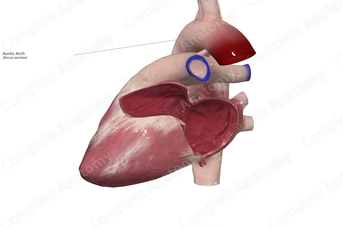
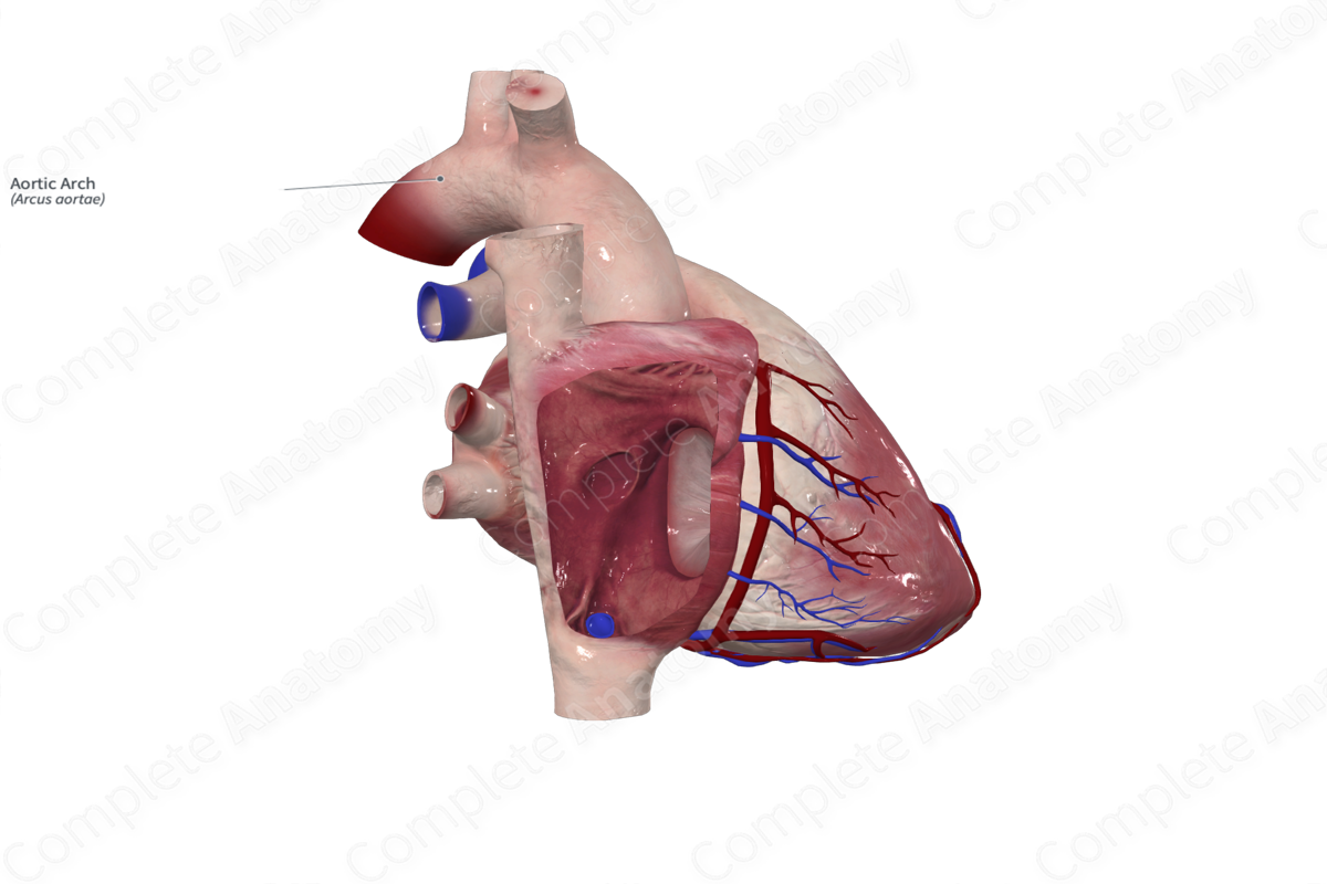
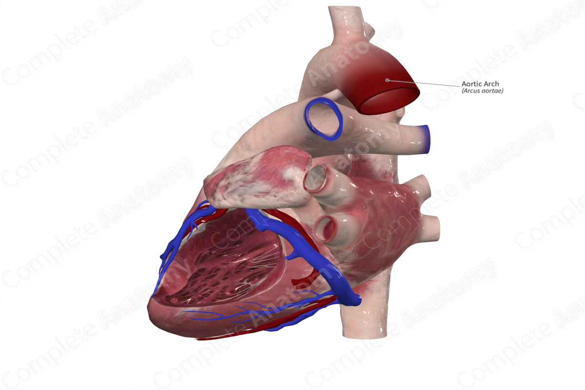
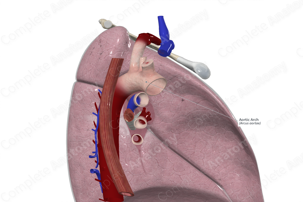
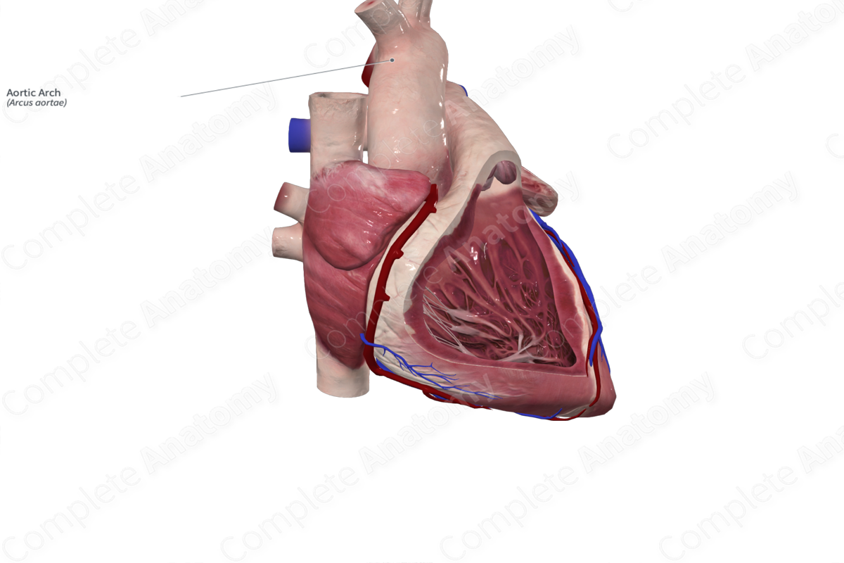
The arch of the aorta is the second part of the aorta and connects the ascending and the descending portions. It originates at the upper margin of the second sternocostal joint on the right side and ascends obliquely in a posterior direction. The superior-most point of the arch is 2.5 cm below the sternal notch. After it peaks, it descends to the left of the fourth thoracic vertebra and becomes the descending aorta at the level of the second costal cartilage. As it lies above the transverse thoracic plane, the arch sits within the superior mediastinum.
Related parts of the anatomy
Key Features/Anatomical Relations
Several nerves cross the arch, including the left phrenic nerve, as it travels towards the pericardium, and the left vagus nerve. The left vagus nerve gives off the left recurrent laryngeal branch after it passes inferior to the aortic arch. This recurrent laryngeal branch wraps around the aorta, posterior to the ligamentum arteriosum, and ascends towards the larynx on the right-hand side of the aortic arch. The ligamentum arteriosum sits inferior to the aortic arch and connects it to the bifurcation of the pulmonary trunk. It’s a remnant of the ductus arteriosus that is found during fetal development.
The superficial cardiac plexus resides between the bifurcation of the pulmonary trunk and the aortic arch, in the region of the ligamentum arteriosum. Between the origins of the brachiocephalic trunk and the left common carotid artery, the left superior intercostal vein is seen as it travels superiorly towards the left brachiocephalic vein. The trachea, esophagus, thoracic duct, and deep cardiac plexus all lie posterior to the arch.
Function
The arch of aorta transmits oxygenated blood from the ascending aorta to the descending aorta, where it’s eventually distributed to the thoracic, abdominal, pelvic, and lower limb regions. It is under considerable pressure during ventricular systole; therefore, has a thick muscular wall and a high elastin content. Additionally, there are three branches of the aortic arch, the brachiocephalic trunk, left common carotid artery, and left subclavian artery. Several anatomical variations have been described for the origin of these vessels on the arch, including the distance between their origins. Some may even share a common trunk (Tubbs, Shoja and Loukas, 2016). These three branches are responsible for delivering oxygenated blood to the head, neck, and upper limb.
List of Clinical Correlates
- Aortic aneurysm
- Aortic dissection
- Coarctation of the aorta
References
Tubbs, R. S., Shoja, M. M. and Loukas, M. (2016) Bergman's Comprehensive Encyclopedia of Human Anatomic Variation. Wiley.
Learn more about this topic from other Elsevier products

