Structure/Morphology
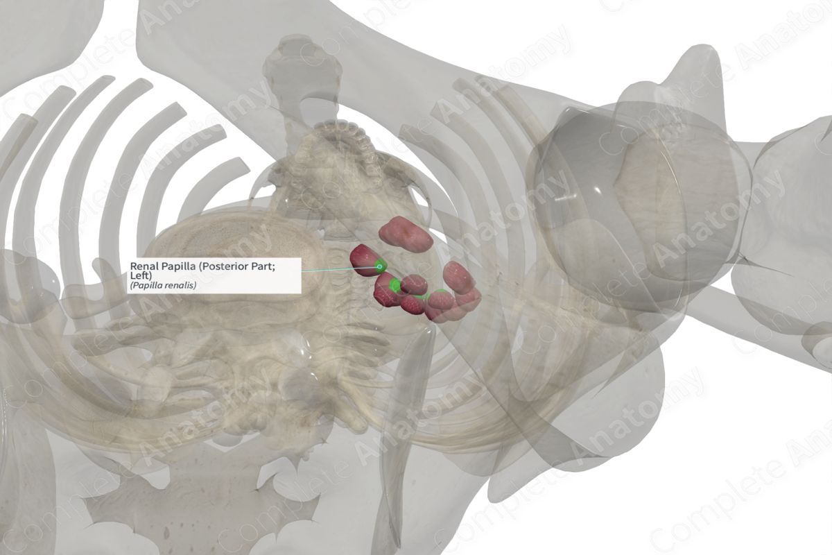
The renal papillae are described as the apex or pointed end of each renal pyramid. Each papilla projects into a minor calix, which lies in the central space of the renal pelvis.
A distinct feature of papillae is their sieve-like appearance, which can be attributed to the presence of many small openings on their surface. Each opening represents a small tubule called the papillary duct, into which the collecting tubules within the renal pyramid converge.
Related parts of the anatomy
Key Features/Anatomical Relations
Renal papillae are located at the apex of the renal pyramids at the junction of the renal medulla and renal pelvis.
Function
Renal papillae carry renal filtrate from the papillary ducts to the minor calices of the renal pelvis via papillary ducts.
List of Clinical Correlates
—Renal papillary necrosis
Learn more about this topic from other Elsevier products


