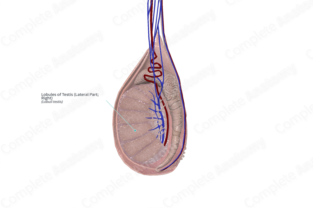
Structure/Morphology
Each testis has approximately 250 lobules separated by septa (Standring, 2016). The lobules are conical in shape, with the base oriented near the circumference of the testis and the apex directed towards the mediastinum.


Key Features/ Anatomical Relations
The lobules consist of one to four seminiferous tubules and interstitial tissue (consisting of blood vessels, nerves, Leydig cells, and macrophages).
Function
The lobules serve as the location for seminiferous tubules (which are responsible for the production of sperm) and Leydig cells (which are responsible for the production of testosterone).
References
Standring, S. (2016) Gray's Anatomy: The Anatomical Basis of Clinical Practice. Gray's Anatomy Series 41 edn.: Elsevier Limited.
Learn more about this topic from other Elsevier products

