Quick Facts
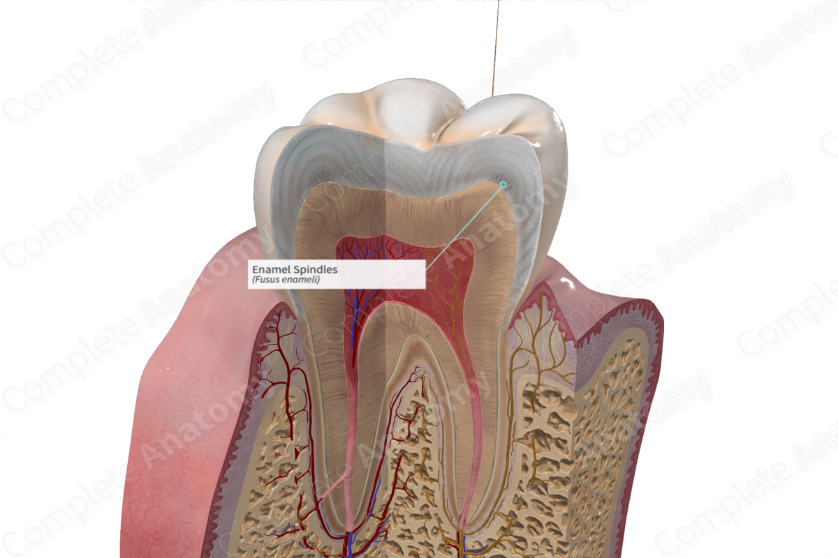
The enamel spindles are the clublike structures in the inner third of the dental enamel, believed to be terminals of protoplasmic processes of the odontoblasts that have passed across the dentinoenamel junction (Dorland, 2011).
Related parts of the anatomy
Structure and/or Key Feature(s)
Enamel spindles are the projections of dentinal tubules that occasionally traverse the dentinoenamel junction and extend into the enamel. Often more prevalent at the cusp tips, these spindles appear as a series of dilated ampulla-shaped extensions anchored to the dentinoenamel junction (Berkovitz et al., 2012).
The spindles are formed during tooth development when odontoblast processes become entrapped between ameloblasts prior to and during amelogenesis (Standring, 2016).
References
Berkovitz, B. K. B., Boyde, A., Frank, R. M., Höhling, H. J., Moxham, B. J., Nalbandian, J. and Tonge, C. H. (2012) Teeth. Springer Berlin Heidelberg.
Dorland, W. (2011) Dorland's Illustrated Medical Dictionary. 32nd edn. Philadelphia, USA: Elsevier Saunders.
Standring, S. (2016) Gray's Anatomy: The Anatomical Basis of Clinical Practice. Gray's Anatomy Series 41 edn.: Elsevier Limited.
