Quick Facts
Location: Vertebral column.
Bone Type: Irregular bone.
Key Features: Vertebral body, laminae, pedicles, superior and inferior articular processes, and transverse and spinous processes.
Articulates With: Eleventh thoracic and first lumbar vertebra, twelfth ribs.
Arterial Supply: Subcostal arteries.


Key Features & Anatomical Relations
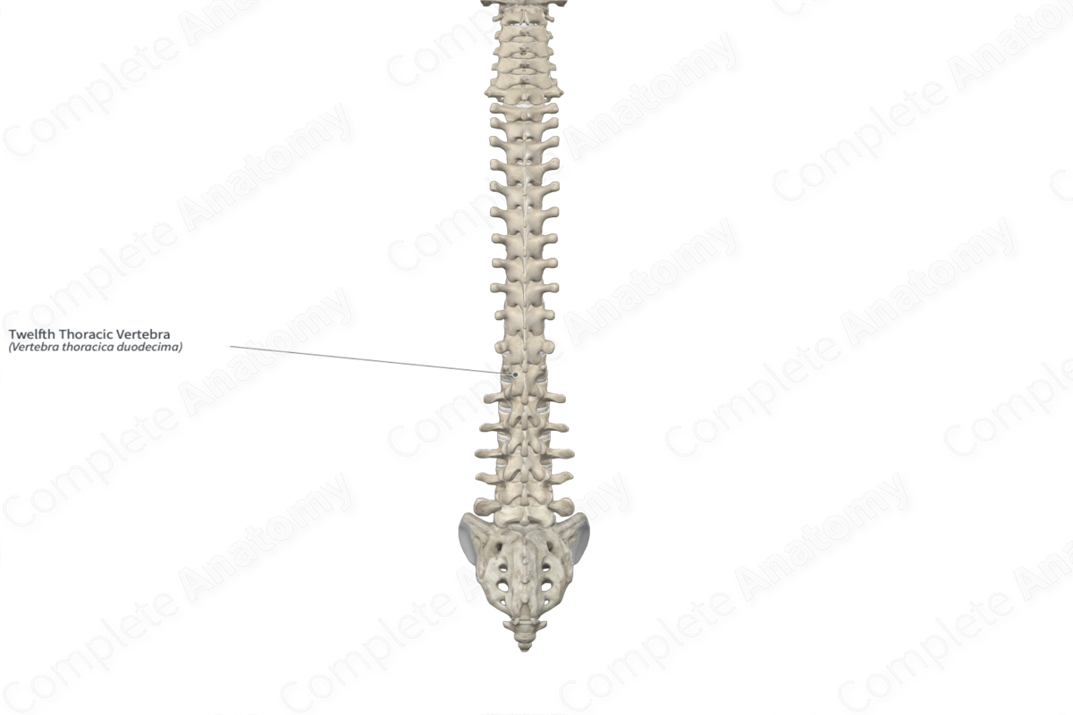
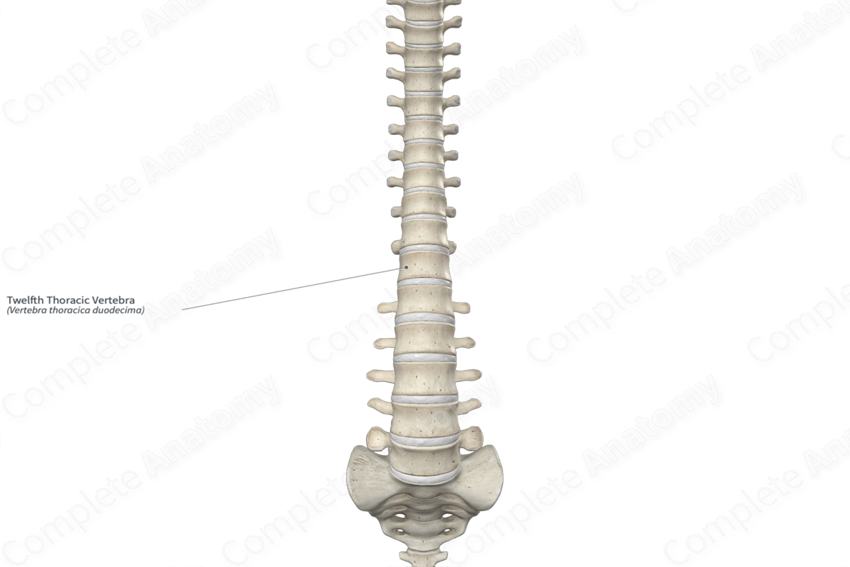
The twelfth thoracic vertebra (vertebra T12) is the largest of the twelve thoracic vertebrae of the vertebral column. Although its general morphology is typical of the thoracic vertebrae, the twelfth thoracic vertebra presents a number of transitionary features, similar to the first lumbar vertebra. It is classified as an irregular bone and includes the following bony features:
- parts: vertebral body, laminae, pedicles, superior and inferior articular processes, and transverse and spinous processes;
- surfaces: superior and inferior intervertebral surfaces, superior and inferior annular epiphyses, and vertebral arch;
- landmarks: superior and inferior vertebral notches, superior and inferior articular facets, costal facets, and superior, inferior and lateral tubercles.
More information regarding these and other bony features can be found in the Parts, Surfaces, and Landmarks tabs for this bone.
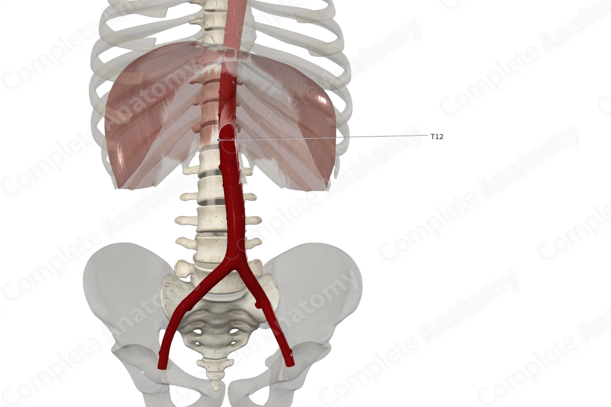
The twelfth thoracic vertebra is located:
- superior to the first lumbar vertebra;
- inferior to the eleventh thoracic vertebra;
- medial to the twelfth rib.
It articulates with the:
- eleventh thoracic and first lumbar vertebrae at the intervertebral symphyses and zygapophyseal joints;
- twelfth rib at the joint of head of twelfth rib.
Ossification
Ossification of all thoracic vertebrae occurs at eight ossification centers, these are found in the:
- vertebral body, which appears in utero during the second to fourth months;
- right and left halves of the vertebral arch, with one center found in each, which appear in utero during the third month;
- right and left transverse processes, with one center found in each, which appear during puberty;
- spinous process, which appears during puberty;
- superior and inferior annular epiphyses, with one center found in each, which appear during puberty.
The ossification centers for the right and left halves of the vertebral arch fuse with each other during the first year after birth. The vertebral arch fuses with the vertebral body during the third year. The remaining centers fuse with the vertebral arch and body during early adulthood (Standring, 2016).
Variations
In some individuals:
- the pedicles of thoracic vertebrae can display significant morphological variation;
- the transverse processes may fail to ossify (Tubbs, Shoja and Loukas, 2016).
Surface Anatomy
The spinous process of the twelfth thoracic vertebra can be palpated, especially during flexion of the neck trunk.
List of Clinical Correlates
- Fracture
- Osteoporosis
- Spinal stenosis
- Scoliosis
- Spondylosis
- Spondylolisthesis
- Spondylolysis
- Butterfly vertebra
- Spina bifida
References
Standring, S. (2016) Gray's Anatomy: The Anatomical Basis of Clinical Practice. Gray's Anatomy Series 41st edn.: Elsevier Limited.
Tubbs, R. S., Shoja, M. M. and Loukas, M. (2016) Bergman's Comprehensive Encyclopedia of Human Anatomic Variation. Wiley.
Learn more about this topic from other Elsevier products

