Quick Facts
Location: Forearm.
Bone Type: Long bone.
Key Features: Head, body, olecranon, trochlear and radial notches, and ulnar styloid process.
Articulates With: Humerus and radius.
Arterial Supply: Anterior and posterior interosseous, radial, and ulnar arteries.


Related parts of the anatomy
Key Features & Anatomical Relations
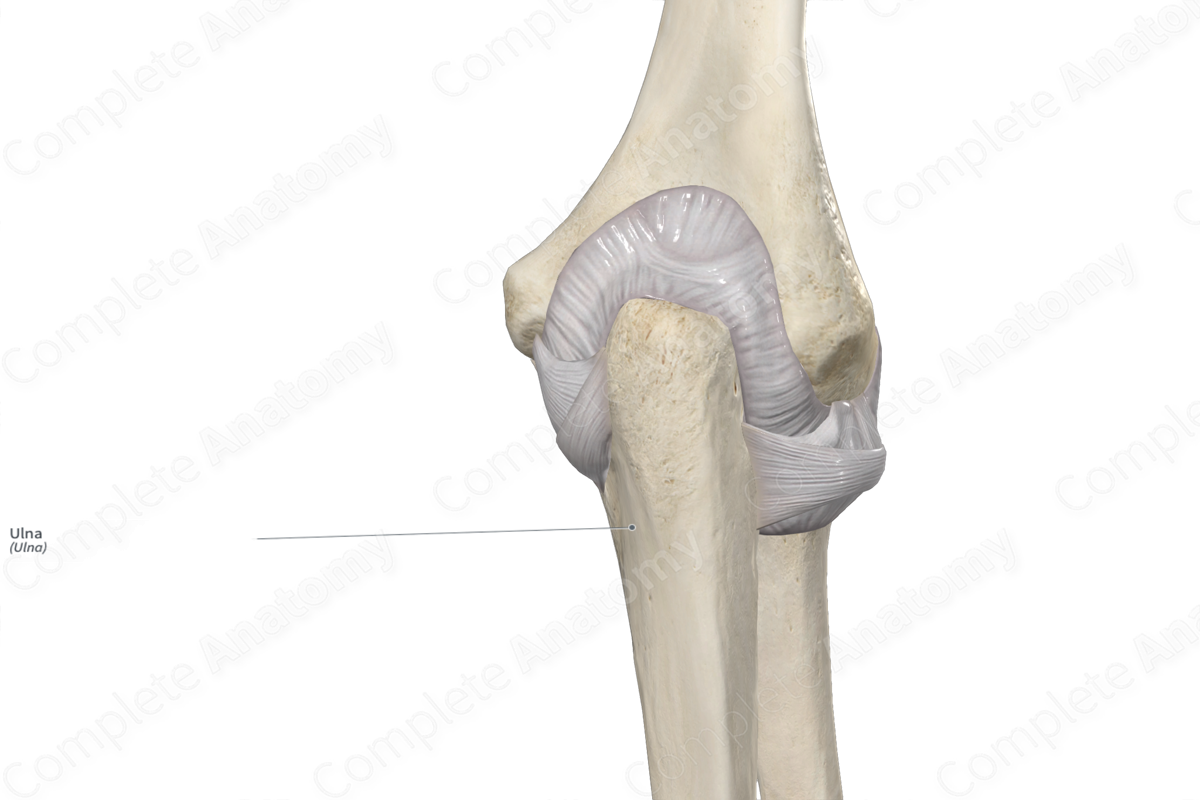
The ulna is the longer of the two bones of the forearm, the other being the radius. It’s classified as a long bone and includes the following bony features:
- parts: proximal part, body, and distal part;
- surfaces: anterior, posterior, and medial surfaces, and anterior, posterior, and interosseous borders;
- landmarks: head, olecranon, trochlear and radial notches, tuberosity of ulna, and ulnar styloid process.
More information regarding these and other bony features can be found in the Parts, Surfaces and Landmarks tabs for this bone.
The ulna is located:
- proximal to the articular disc of the distal radioulnar joint;
- distal to the humerus;
- medial to the radius.
It articulates with the:
- humerus at the humeroulnar joint, contributing to the formation of the elbow joint;
- radius at both the proximal and distal radioulnar joints.
Unlike the radius, the ulna does not articulate with any of the carpal bones but is instead separated from them by the articular disc of the distal radioulnar joint.
Ossification
Ossification of the ulna occurs at four ossification centers, these are found in the:
- body, which appears in utero at the eighth week;
- proximal part, two centers are located here and appear within the ninth to eleventh years;
- distal part, which appears within the fifth to sixth years.
The ossification center for the proximal part fuses with the body within the fourteenth to eighteenth years, while the ossification center for the distal part of ulna fuses with the body within the seventeenth to eighteenth years (Standring, 2016).
Variations
In some individuals, interrupted growth of the epiphyses of the ulna can result in hypoplasia, and partial or complete aplasia of the bone (Tubbs, Shoja and Loukas, 2016).
Surface Anatomy
The following bony features of the ulna are relevant to surface anatomy:
- the olecranon is subcutaneous and therefore, when the forearm is flexed at the elbow joint, the olecranon forms the apex on the posterior aspect of the joint;
- the posterior border is subcutaneous and can be palpated along its entire length;
- the head is located by palpating the medial aspect of the wrist joint;
- the ulnar styloid process is easily identified by the large prominence on the medial side of the dorsal aspect of the wrist.
List of Clinical Correlates
- Fracture of ulna (e.g., Monteggia fracture)
- Radioulnar synostosis
References
Standring, S. (2016) Gray's Anatomy: The Anatomical Basis of Clinical Practice. Gray's Anatomy Series 41st edn.: Elsevier Limited.
Tubbs, R. S., Shoja, M. M. and Loukas, M. (2016) Bergman's Comprehensive Encyclopedia of Human Anatomic Variation. Wiley.
Learn more about this topic from other Elsevier products

