Quick Facts
Location: Forearm.
Bone Type: Long bone.
Key Features: Head, neck, body, radial tuberosity, radial styloid process, and ulnar notch.
Articulates With: Humerus, ulna, scaphoid bone, and lunate bone.
Arterial Supply: Anterior and posterior interosseous, radial, and ulnar arteries.


Related parts of the anatomy
Key Features & Anatomical Relations
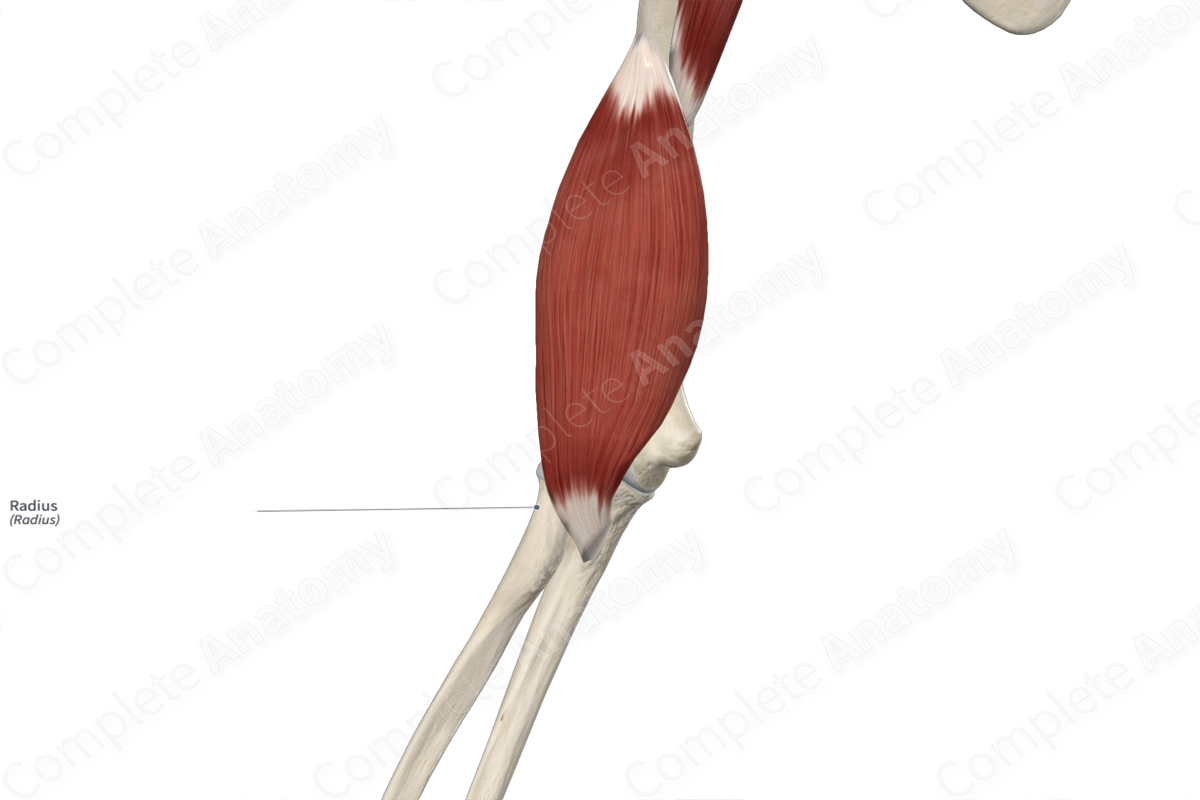
The radius is the shorter of the two bones of the forearm, the other being the ulna. The name “radius” is derived from its ability to rotate around the more fixed ulna. It’s classified as a long bone and includes the following bony features:
- parts: proximal part, body and distal part;
- surfaces: anterior, posterior and lateral surfaces, and anterior, posterior and interosseous borders;
- landmarks: head, neck, radial tuberosity, radial styloid process, dorsal radial tubercle, and ulnar notch.
More information regarding these and other bony features can be found in the Parts, Surfaces and Landmarks tabs for this bone.
The radius is located:
- proximal to the scaphoid and lunate bones;
- distal to the humerus;
- lateral to the ulna.
It articulates with the:
- humerus at the humeroradial joint, contributing to the formation of the elbow joint;
- ulna at both the proximal and distal radioulnar joints;
- scaphoid bone, at the radioscaphoid joint, contributing to the formation of the radiocarpal (wrist) joint;
- lunate bone, at the radiolunate joint, also contributing to the formation of the radiocarpal joint.
Ossification
Ossification of the radius occurs at three ossification centers, these are found in the:
- body, which appears in utero at the eighth week;
- proximal part, which appears within the fourth to fifth years;
- distal part, which appears within the first year.
The ossification center for the proximal part of radius fuses with the body within the fourteenth to eighteenth years. The ossification center for the distal part of radius fuses with the body within the seventeenth to nineteenth years (Standring, 2016).
Variations
In some individuals, an accessory tubercle, known as a lunar tubercle, may be present at the distal part of radius (Tubbs, Shoja and Loukas, 2016).
Surface Anatomy
The following bony features of the radius are relevant to surface anatomy:
- the head can be located distal to the lateral epicondyle of the humerus in the extended elbow joint;
- the radial styloid process can be felt by palpation of the anatomical snuff box;
- proximal to the radial styloid process, the anterior, posterior and lateral surfaces are subcutaneous and easily palpable;
- the dorsal radial tubercle can be palpated on the dorsolateral aspect of the wrist.
List of Clinical Correlates
- Dislocation of elbow joint
- Radioulnar synostosis
- Radial dysplasia
- Fracture of radius (e.g., Colles’, Smith’s, Barton’s, and Galeazzi fractures)
- Madelung’s deformity
References
Standring, S. (2016) Gray's Anatomy: The Anatomical Basis of Clinical Practice. Gray's Anatomy Series 41st edn.: Elsevier Limited.
Tubbs, R. S., Shoja, M. M. and Loukas, M. (2016) Bergman's Comprehensive Encyclopedia of Human Anatomic Variation. Wiley.
Learn more about this topic from other Elsevier products

