Quick Facts
Location: Pelvic girdle.
Bone Type: Flat bone.
Key Features: Body, ala, iliac crest, iliac fossa, and anterior superior iliac spine.
Articulates With: Ischium, pubis, sacrum, and femur.
Arterial Supply: Iliolumbar, obturator, superficial circumflex iliac, and superior gluteal arteries.
Related parts of the anatomy
Key Features & Anatomical Relations
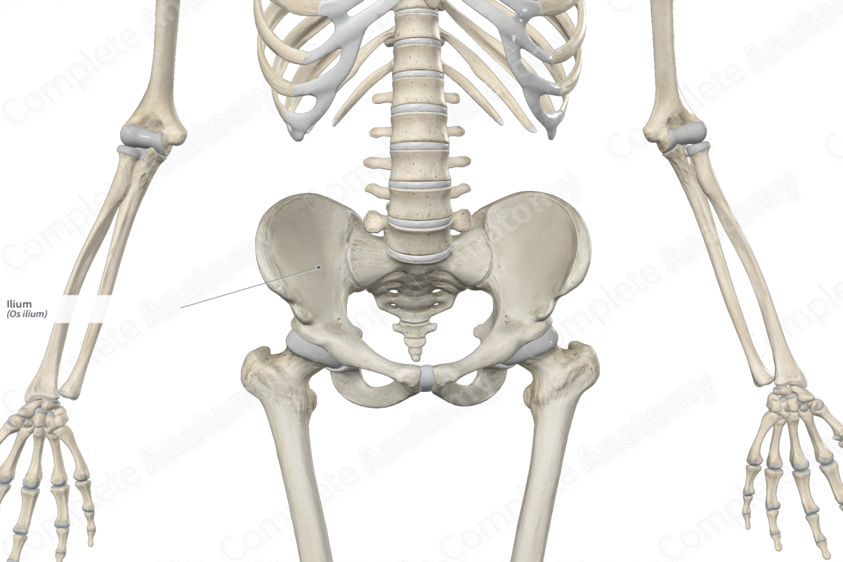
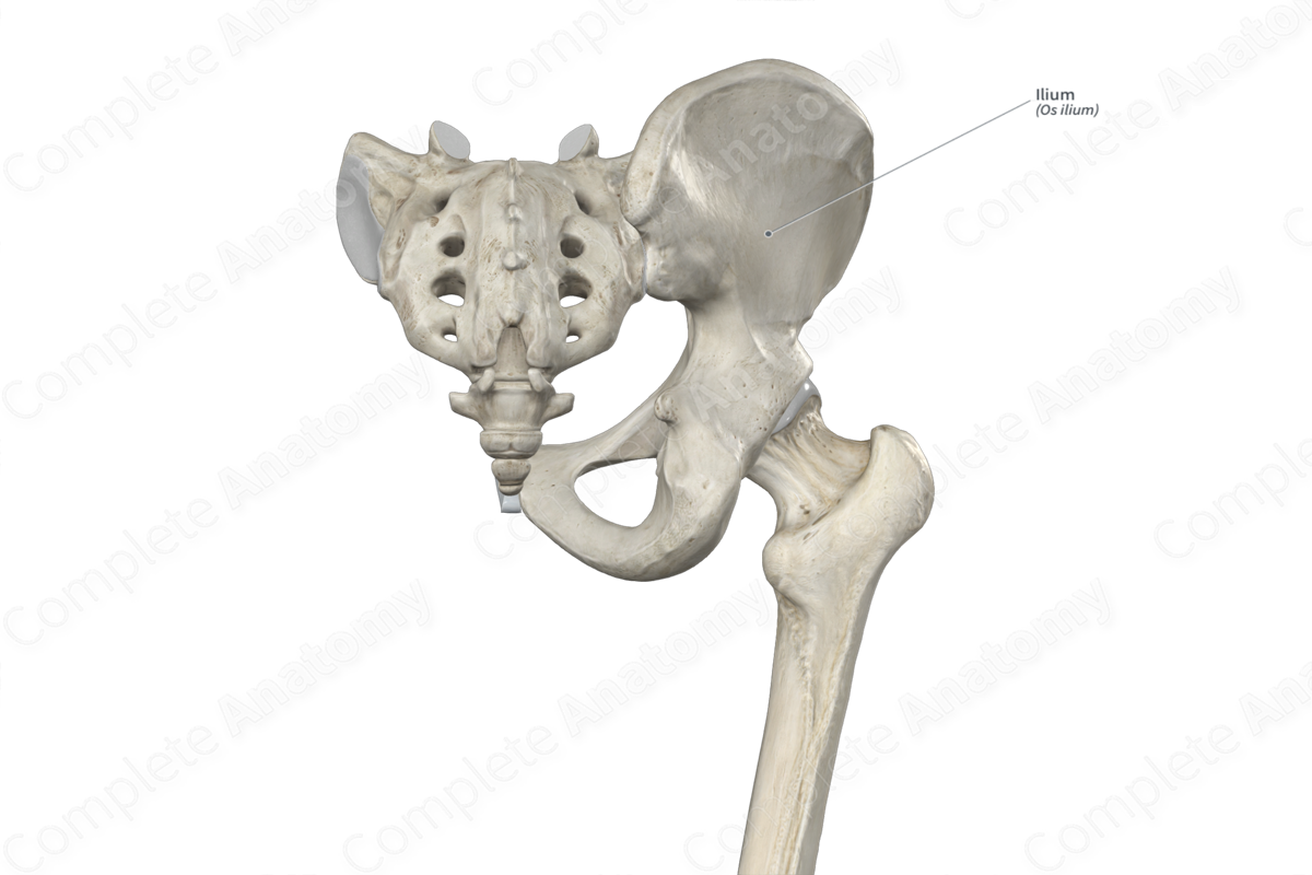
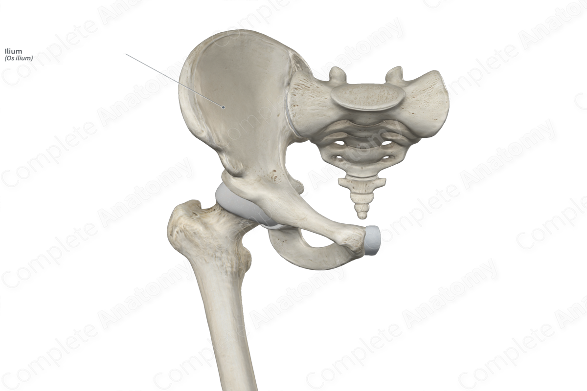
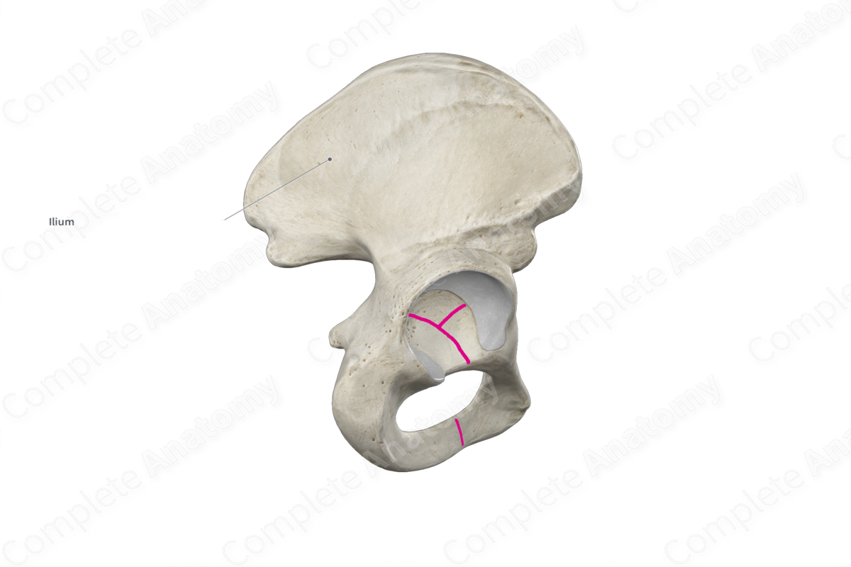
The ilium (or iliac bone) is the largest of the three bones that form the hip bone, the other two being the ischium and pubis. It’s a large, fan-like bone that is found along the superolateral aspect of the pelvic girdle. The ilium is classified as a flat bone and includes the following bony features:
- parts: body and ala;
- surfaces: gluteal and sacropelvic surfaces, anterior, posterior and medial borders, and iliac fossa;
- landmarks: anterior superior and posterior superior iliac spines, anterior, posterior and inferior gluteal lines, and iliac tubercle.
More information regarding these bony features can be found in the Parts, Surfaces and Landmarks tabs for this bone.
The ilium is located:
- proximal to the femur;
- superior to the ischium and pubis;
- lateral to the sacrum and fifth lumbar vertebra.
It articulates with the:
- ischium and pubis, via triradiate cartilage in childhood and fused joints in adulthood;
- sacrum at the sacroiliac joint;
- femur at the hip joint.
Ossification
Ossification of the ilium occurs at three ossification centers, these are found in the:
- body of ilium, which appears in utero at the ninth week;
- iliac crest, two centers are located here and appear within early to middle adolescence.
These ossification centers fuse with each other during middle adolescence to early adulthood. Fusion between the body of ilium, body of ischium, and superior pubic ramus occurs at the acetabulum during middle to late adolescence, forming the fused hip bone (Standring, 2016).
Variations
The ilium displays sexual dimorphism, where:
- the iliac crest tends to be more rugged and curved in males;
- the ala of ilium tends to be more sloped and less vertical in males.
Surface Anatomy
The following bony features of the ilium are relevant to surface anatomy:
- the anterior one third of the iliac crest is subcutaneous and therefore easily palpated;
- the anterior superior iliac spine can be located by following the iliac crest to its anterior end point;
- the iliac tubercle is palpable along the iliac crest, approximately 5–6 cm posterior to the anterior superior iliac spine.
List of Clinical Correlates
- Fracture of ilium
- Iliac bone graft harvesting
- Iliac horn syndrome
References
Standring, S. (2016) Gray's Anatomy: The Anatomical Basis of Clinical Practice. Gray's Anatomy Series 41st edn.: Elsevier Limited.
