Quick Facts
Location: Hand.
Bone Type: Short bone.
Key Features: Hook, palmar and dorsal surfaces; triquetrum, capitate, and lunate articular facets.
Articulates With: Capitate, lunate, triquetrum, and fourth and fifth metacarpal bones.
Arterial Supply: Deep palmar arch, dorsal carpal branch of radial and ulnar arteries.
Related parts of the anatomy
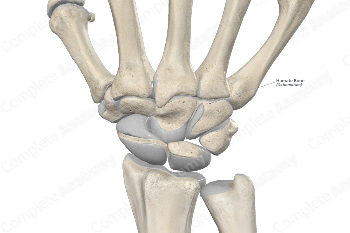
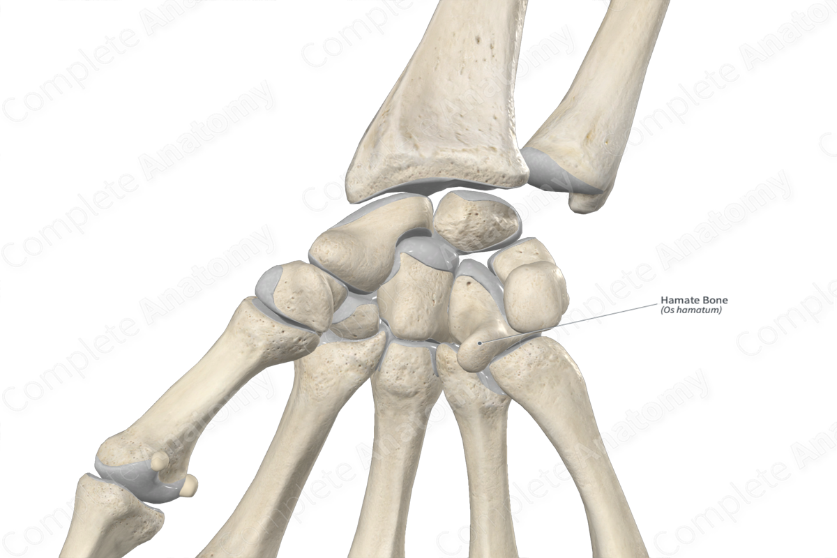
Key Features & Anatomical Relations
The hamate bone (or unciform bone) is one of the eight carpal bones of the hand. It’s wedge-shaped, has a characteristic hook (hamulus), and is found in the distal row of carpal bones. It’s classified as a short bone and includes the following bony features:
- parts: hook;
- surfaces: palmar and dorsal surfaces;
- landmarks: triquetrum, capitate and lunate articular facets, and fourth and fifth metacarpal articular facets.
More information regarding these bony features can be found in the Parts, Surfaces and Landmarks tabs for this bone.
The hamate bone is located:
- proximal to the fourth and fifth metacarpal bones;
- distal to the lunate bone;
- medial to the capitate bone;
- lateral to the triquetrum bone.
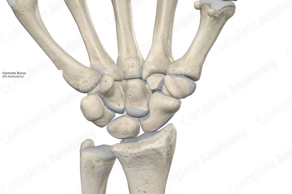
It articulates with the:
- capitate bone at the capitohamate joint;
- lunate bone at the lunohamate joint;
- triquetrum bone at the triquetrohamate joint;
- fourth and fifth metacarpal bones, contributing to the formation of the carpometacarpal joints.
Ossification
Ossification of the hamate bone occurs at one ossification center, which appears within the third month after birth (Standring, 2016). Complete ossification occurs during middle to late adolescence.
Variations
In some individuals:
- carpal fusion, or coalition, between the hamate and capitate bones may be present, forming the capitohamate bone;
- several accessory bones may be associated with the hamate bone (Tubbs, Shoja and Loukas, 2016).
Surface Anatomy
With regard to surface anatomy, the hook of hamate bone can be palpated between the hypothenar eminence and the pisiform bone.
List of Clinical Correlates
- Fracture of hamate bone (common in golfers, baseball and tennis players)
References
Standring, S. (2016) Gray's Anatomy: The Anatomical Basis of Clinical Practice. Gray's Anatomy Series 41st edn.: Elsevier Limited.
Tubbs, R. S., Shoja, M. M. and Loukas, M. (2016) Bergman's Comprehensive Encyclopedia of Human Anatomic Variation. Wiley.
Learn more about this topic from other Elsevier products

