Quick Facts
Location: Thigh.
Bone Type: Long bone.
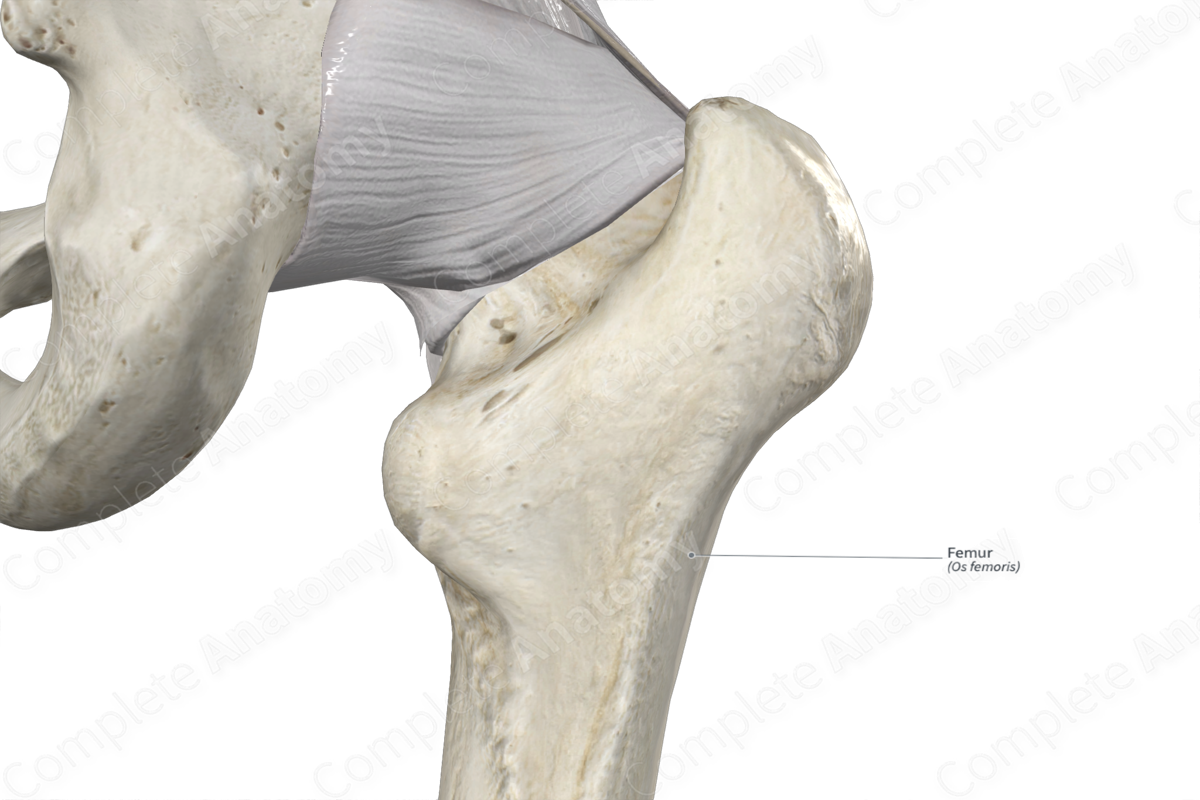
Key Features: Head, neck, body, greater and lesser trochanters, and medial and lateral condyles.
Articulates With: Hip bone, tibia, and patella.
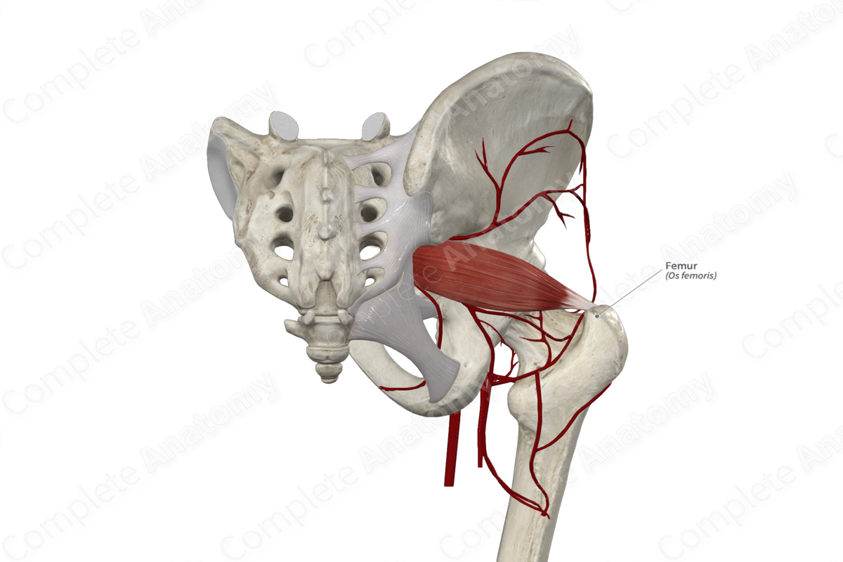
Arterial Supply: Medial and lateral circumflex femoral arteries, superior and inferior gluteal arteries, perforating branches of deep femoral artery, genicular anastomosis.
Related parts of the anatomy
Key Features & Anatomical Relations
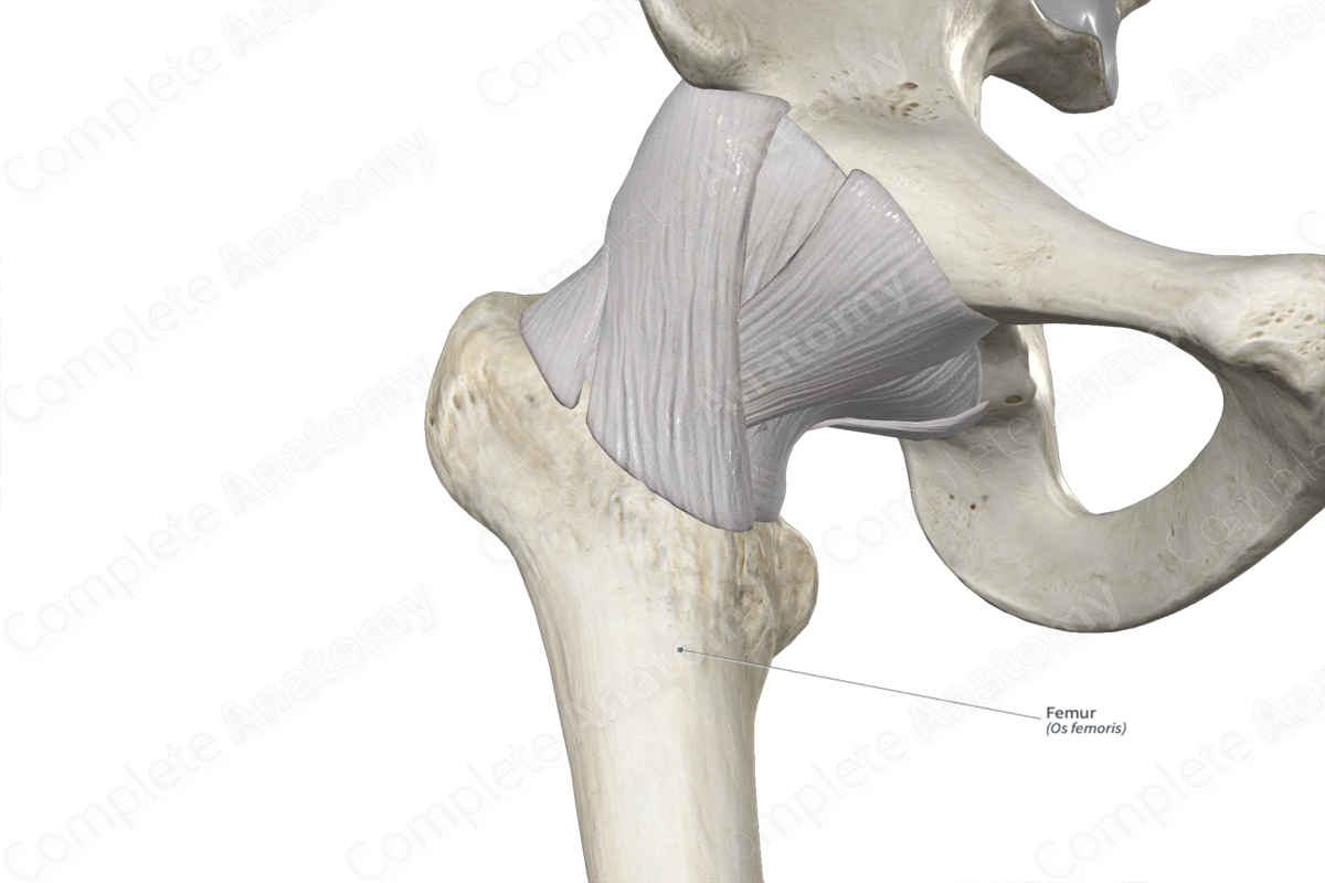
The femur is the single bone found in the thigh and is the longest, heaviest, and strongest bone in the human body. It’s classified as a long bone and includes the following bony features:
- parts: head, neck, body, greater and lesser trochanters, and medial and lateral condyles;
- surfaces: anterior, posterior, medial and lateral surfaces, medial and lateral borders, and linea aspera;
- landmarks: trochanteric fossa, quadrate and adductor tubercles, medial and lateral epicondyles, and pectineal line.
More information regarding these and other bony features can be found in the Parts, Surfaces and Landmarks tabs for this bone.
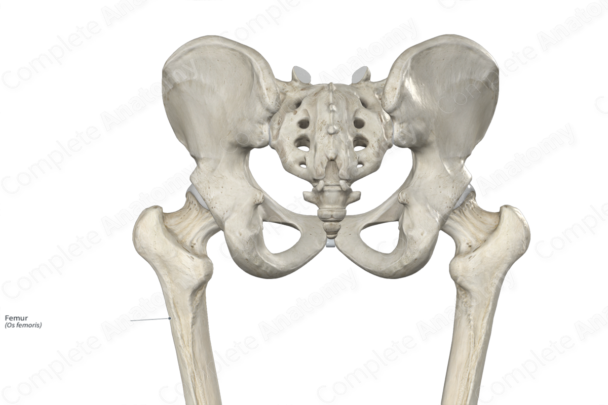
The femur is located:
- distal to the hip bone;
- proximal to the tibia and fibula.
It articulates with the:
- hip bone at the hip joint;
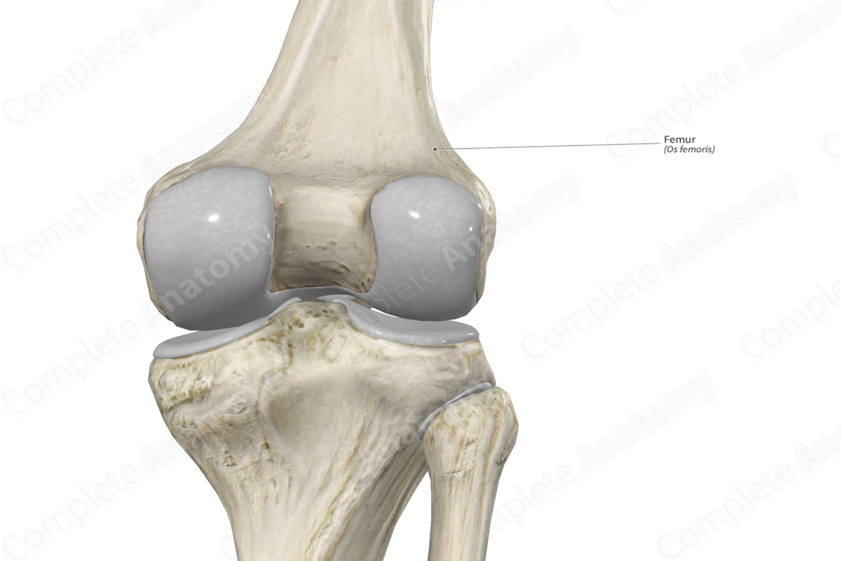
- tibia at the medial and lateral femorotibial joints, contributing to the formation of the knee joint;
- patella at the patellofemoral joint, also contributing to the formation of the knee joint.
Ossification
Ossification of the femur occurs at five ossification centers, these are found in the:
- body, which appears in utero during the second month;
- head, which appears within the first six months after birth;
- greater trochanter, which appears during the fourth year;
- lesser trochanter, which appears within the twelfth to fourteenth years;
- distal part of femur, which appears during the ninth month after birth.
The ossification centers for the head, greater trochanter, lesser trochanter, and distal part of femur fuse with the body during middle to late adolescence (Standring, 2016).
Variations
The distal part of femur presents several variations of shape and size depending on ethnicity and gender. In some individuals:
- the body of the femur may have a third nutrient foramen;
- the linea aspera may present with an underlying bony elevation, known as linea aspera-pilaster complex;
- the articular facets for the medial and lateral condyles of the femur may present with transversely oriented grooves, which receive portions of the medial and lateral menisci during knee extension, respectively (Tubbs, Shoja and Loukas, 2016).
Surface Anatomy
The following bony features of the femur are relevant to surface anatomy:
- the head can be palpated inferior to the midpoint of the inguinal ligament;
- the greater trochanter can be palpated anterolateral to the buttocks;
- the medial and lateral condyles are subcutaneous and therefore easily identified, especially during flexion of the knee joint;
- the medial and lateral epicondyles are very prominent and easily palpated;
- the patellar surface of the femur is palpable during flexion of the knee joint.
List of Clinical Correlates
- Fracture of femur (e.g. femoral neck, trochanteric, mid-shaft, and distal fractures)
- Coxa vara/valga
- Osteoarthritis of hip joint
- Total hip arthroplasty
References
Standring, S. (2016) Gray's Anatomy: The Anatomical Basis of Clinical Practice. Gray's Anatomy Series 41st edn.: Elsevier Limited.
Tubbs, R. S., Shoja, M. M. and Loukas, M. (2016) Bergman's Comprehensive Encyclopedia of Human Anatomic Variation. Wiley.
